SLIDE 1 Janet Iwasa
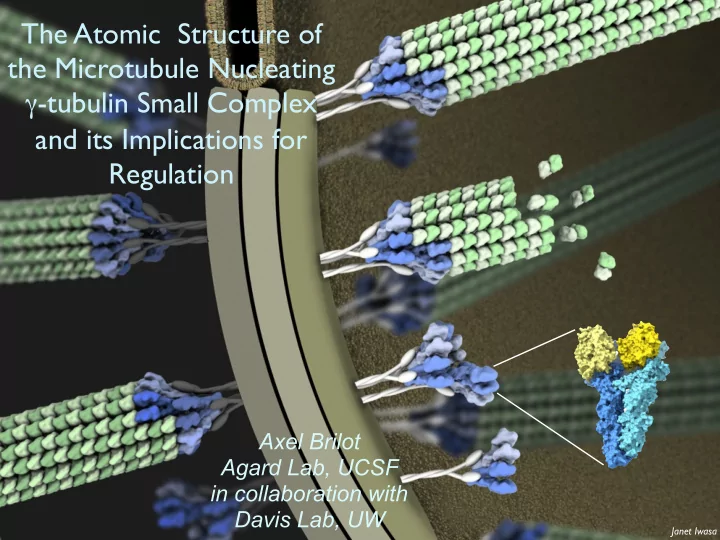
Axel Brilot Agard Lab, UCSF in collaboration with Davis Lab, UW
The Atomic Structure of the Microtubule Nucleating γ-tubulin Small Complex and its Implications for Regulation
SLIDE 2 Cryo-EM tomogram / Sam Li
Yeast Spindle
Spindle Pole Body (yeast centrosome) Spc110p γTuRC Microtubule
Microtubule nucleation by γ-tubulin Complexes
γTuSC/γTuRC structure, assembly, activation
γ-Tubulin Small Complex (γTuSC) 2 γ-Tubulins, GCP2,GCP3 (Spc97 Spc98)
GPC3 G P C 2
γ γ
SLIDE 3 Individual γΤuSCs
50 nm
GPC3 G P C 2
γ γ
+ Spc110p1-220
γTuRCs/filaments
100 nm
γTuRC
Filament
6.5 γTuSCs/turn = 13 γ-tubulins = in vivo MT protofilament #
90º
Attachment factor Spc110 stabilizes γTuSC assembly
spindle pole body
Spc110p
SLIDE 4 Spc110
Model: γTuRC nucleating microtubule intra-γTuSC inter-γTuSC
Open state
intra-γTuSC inter-γTuSC
Closed state (disulfide stabilized)
Open - closed transition enhances γ-TuRC MT nucleation
- J. Kollman
- closed state better MT nucleator
- suggests closure as a regulatory mechanism
SLIDE 5 γTuSC pseudo-atomic model built using 6.5Å oxidized map
filament interior
GCP2 GCP3 γ-tubulin
- C. Greenberg/A. Sali
- J. Kollman
Merdes, Mourney Guillet, et al. 2011
GCP4 crystal structure
C-terminal domain directly binds γ-tubulin
less than 20% similarity
SLIDE 6 γTuSC pseudo-atomic model built using 6.5Å oxidized map
filament interior
GCP2 GCP3 γ-tubulin
- C. Greenberg/A. Sali
- J. Kollman
Merdes, Mourney Guillet, et al. 2011
GCP4 crystal structure
C-terminal domain directly binds γ-tubulin
less than 20% similarity
Missing 234 aa from gcp2, 275 aa from gcp3 Built into a ~6.5 Å map
SLIDE 7 Polara Data
~80 e-/A2 Dose filtered & aligned with MotionCorr2 Thon rings 5Å
The Image Data
SLIDE 8 γTuSC monomer/dimer by single particle cryoEM (3.8Å)
- first true atomic description of γTuSC, numerous inserts, etc
- differences in the interfaces between the γTuSCs vs internal interface
- conformational changes in γ-tubulin upon assembly into γTuSC
- interpretation of phosphorylation sites, mutations
SLIDE 9
Workflow
Drift correct & pick Determine CTF extract particles 2D Classification 3D Classification Extract classes Align into one class 3D Classification
SLIDE 10
Improving the Map
Increase Dataset size (+0.5M particles) Various Programs (Relion, cryosparc) Full workflow, as well as feeding them classification results from Frealign Focused Classification in Frealign Various Masks Half-Tusc, Base only, Base plus one tubulin arm
SLIDE 11
Improving the Map - Frealign, Shaped Masks and Weighting
SLIDE 12
Assembly driven global conformation changes
both assembly & allosteric conformational changes required
SLIDE 13
Assembly driven global conformation changes
both assembly & allosteric conformational changes required
SLIDE 14
Assembly driven global conformation changes
Twist of the conserved GCP domains is the major re-arrangement GCP3 N-terminus Monomer Closed Open
SLIDE 15 What is the conformation of γ-tubulin on the γ-TuSC straight β 98-bound γ Human γ xtal (3CB2)
yeast MT from Nogales & Rice
SLIDE 16
What is the conformation of γ-tubulin on the γ-TuSC straight α straight β 98-bound γ Human γ xtal (3CB2)
SLIDE 17
What is the conformation of γ-tubulin on the γ-TuSC clashes w/human γ-tubulin clashes with human and yeast γ-tubulin straight α straight β 98-bound γ Human γ xtal (3CB2)
SLIDE 18 What is the conformation of γ-tubulin on the γ-TuSC straight α straight β 98-bound γ Human γ xtal (3CB2)
species specific conserved
(assembly)
SLIDE 19 GCP2 phospho sites suggests functional roles
GCP2 GCP3 Spc110
γ-tubulin
new phos
SLIDE 20 GCP2 phospho sites suggests functional roles
Spc110 binding
GCP2 GCP3 Spc110
γ-tubulin
new phos
γTuSC recruitment
SLIDE 21 γTuSC assembly
GCP2 phospho sites suggests functional roles
Spc110 binding
GCP2 GCP3 Spc110
γ-tubulin
new phos
SLIDE 22 attachment => assembly
Models for γ-complex mediated attachment and nucleation
MTOC MTOC
γ-tubulin “poised” for α-tubulin binding partially active for nucleation
closure & activation MT nucleation
?
fully active for nucleation
SLIDE 23 Acknowledgements
UCSF
Agard lab Centrosome/MT team David Agard Rose Citron Andrew Lyon Michelle Moritz Sam Li Ray Wang Mariano Tabios EM Core Michael Braunfeld Alex Myasnikov David Bulkley Cameron Kennedy Matthew Harrington Andrej Sali Lab Charles Greenberg Shruthi Viswanath
Beyond
Davis Lab, Univ. of Washington Trisha Davis Eric Muller Tamira Vojnar Genevieve Morin King Yabut Kim Fong Alex Zelter Richard Johnson Connie Peng/David Drubin UCB SPB PO1 Group Mark Winey Trisha Davis Chip Asbury Ivan Rayment Andrej Sali Sue Jasperson