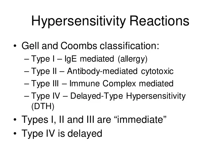

Hypersensitivity Reactions • Gell and Coombs classification: – Type I – IgE mediated (allergy) – Type II – Antibody-mediated cytotoxic – Type III – Immune Complex mediated – Type IV – Delayed-Type Hypersensitivity (DTH) • Types I, II and III are “immediate” • Type IV is delayed
Type I Hypersensitivity • Antigens are called “allergens” • Unknown why people get allergies, but there is a strong genetic predisposition (called atopy) • Hallmark is inappropriate production of IgE against allergens that cause mast cell degranulation (see fig 15-2) • Normally IgE/mast cell activity should be directed against parasitic infections
Type I Hypersensitivity • Mediators of Type I hypersensitivites – Mast cell granule contents (early effects) • Histamine and Heparin - ↑ vascular permeability, smooth muscle contraction (intestines, bronchi), mucus secretion • Chemotactic factors – attract eosinophils and neutrophils • Proteases – mucus secretion, complement activation, degradation of blood vessel basement membrane – Later Effects • Leukotrienes and prostaglandins – secreted after tissue disruption caused by mast cell degranulation, effects are similar to histamine • Arrival of proinflammatory eosinophils and neutrophils
Clinical Manifestations of Type I • Systemic anaphylaxis – Allergen gets into the blood stream – Dyspnea, ↓ BP, bronchole constriction, GI and bladder smooth muscle contration, shock, death within minutes if untreated – Treatment - epinephrine • Allergic rhinitis (hay fever) – Inhaled allergen triggers reaction in nasal mucosa – Watery exudate from nose, eyes, upper respiratory tract, sneeezing and coughing
Clinical Manifestations of Type I • Asthma – Allergic asthma – due to inhaled airborne allergens (pollens, dust, fumes, etc) – Intrinsic asthma – triggered by cold, exercise – Reaction develops in lower respiratory tract – Bronchoconstriction, airway edema, mucus secretion, inflammation • Food allergies – Ingestion of allergen – Vomiting and diarrhea – If allergens are absorbed into bloodstream, reactions can occur where allergen deposits • asthma-like symptoms • Urticaria (hives, wheal & flare response)
Clinical Manifestations of Type I • Atopic Dermatitis (allergic eczema) – Often occurs in young children – Red skin rash – Strong hereditary predisposition
Type I Hypersensitivity • Skin testing – Potential allergens are injected or scratched into the skin – If the patient is allergic a wheal & flare response occurs • RIST – radioimmunosorbent test – similar to RIA, non- invasive way to identify allergies
Type I Hypersensitivity • Treatment – Avoid allergen if possible – Antihistamines, or anti-prostaglandins – Hyposensitization – injections of low doses of allergen may cause a shift from IgE to IgG as the dominant antibody formed.
Type II Hypersensitivity • Antibody-mediated Cytotoxic HS – Antibodies (IgM or IgG) bind to cell surface antigens. Antigen/antibody complex may lead to: • Complement activation lysis • ADCC • Opsonization phagocytosis – These are normal reactions, but when they cause unwarranted tissue damage, they are considered a hypersensitivity.
Type II Hypersensitivity • Examples of Type II HS: – Transfusion reactions • To ABO blood groups • To other RBC blood groups – Hemolytic disease of the newborn (erythroblastosis fetalis) – Drug-induced hemolytic anemia (penicillin)
Type III Hypersensitivity • Immune Complex Disease – Antibody (IgG) / attaching to soluble antigen leads to complex formation – Immune complexes may deposit in: • Blood vessel walls (vasculitis) • Synovial joints (arthritis) • Glomerular basement membrane (glomerulonephritis) • Choroid plexus
Type III Hypersensitivity • Damage occurs due to: – Anaphylatoxin release due to complement activation (C3a, C5a) which then attracts neutrophils, and causes mast cell degranulation – Neutrophils have trouble phagocytosing “stuck” immune complexes so they release their granule contents leading to more inflammation – Platelet aggregation also results from complement activation • These effects are also known as the Arthus reaction
Type III Hypersensitivity • Localized reactions – edema and redness (erythema) and tissue necrosis of the affected tissue – Can occur in the skin following insect bites – Can occur in the lungs • E.g. “farmer’s lung” from inhaling particles from moldy hay
Type III Hypersenstivity • Generalized reactions: – Serum sickness (following treatment with antiserum to a toxin) – Autoimmune diseases • SLE • Rheumatoid arthritis – Drug reactions (penicillin) – Infectious diseases • Meningitis, hepatitis, malaria, mono etc.
Type IV Hypersensitivity • Delayed type hypersensitivity (DTH) – T H cells that have been “sensitized” by an antigen develop a T H 1 and (sometimes a T C response) leading to macrophage recruitment and activation. – First noticed with reaction to tuberculosis bacteria (tuberculin reaction) – Hallmarks of type IV is the large number of macrophages at the reaction site, and that it takes an average of 24 hrs to manifest after repeat exposure.
Recommend
More recommend