
SLIDE 1
9/17/2008 1
Hypersensitivity Hypersensitivity
Stephen Canfield
Assistant Professor Division of Pulmonary, Allergy, and Critical Care Medicine
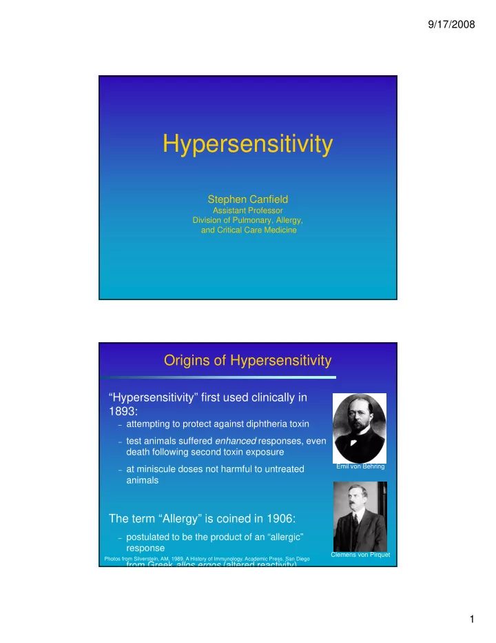
Origins of Hypersensitivity
“Hypersensitivity” first used clinically in 1893:
– attempting to protect against diphtheria toxin – test animals suffered enhanced responses, even
death following second toxin exposure
– at miniscule doses not harmful to untreated
animals
Emil von Behring
The term “Allergy” is coined in 1906:
– postulated to be the product of an “allergic”
response
– from Greek allos ergos (altered reactivity)
Photos from Silverstein, AM. 1989. A History of Immunology. Academic Press, San Diego
Clemens von Pirquet