
SLIDE 1
1
Hypersensitivity
Stephen Canfield, MD PhD
Assistant Professor Division of Pulmonary, Allergy and Critical Care Medicine
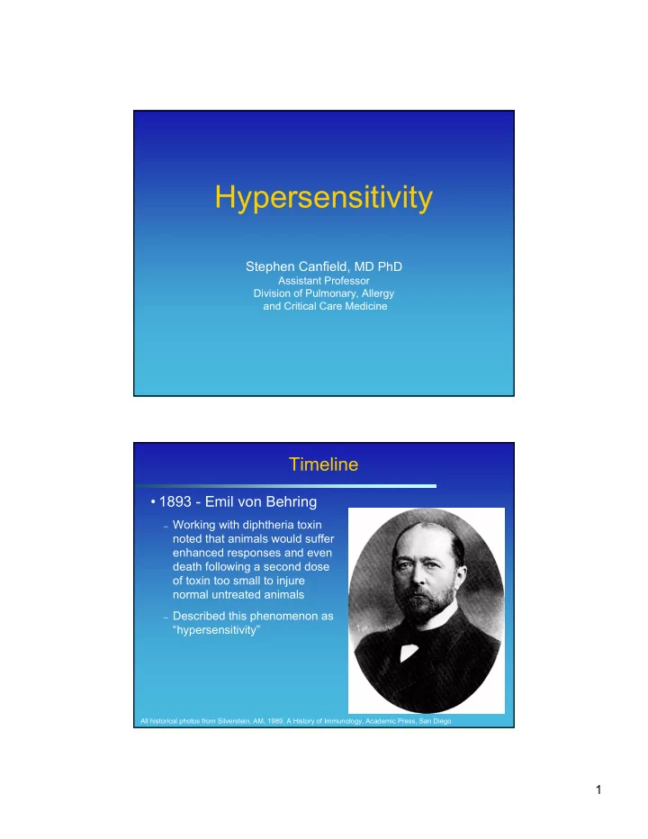
Timeline
- 1893 - Emil von Behring
– Working with diphtheria toxin
noted that animals would suffer enhanced responses and even death following a second dose
- f toxin too small to injure
normal untreated animals
– Described this phenomenon as
“hypersensitivity”
All historical photos from Silverstein, AM. 1989. A History of Immunology. Academic Press, San Diego