SLIDE 1
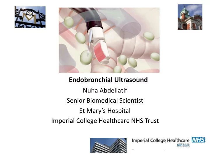
Endobronchial Ultrasound
Nuha Abdellatif Senior Biomedical Scientist St Mary’s Hospital Imperial College Healthcare NHS Trust
SLIDE 2
SLIDE 3 Expanding beyond the airway
- Airway lesions visible by conventional bronchoscopy
- Mediastinal node sampling
- Peri-bronchial lesions not visible
SLIDE 4
SLIDE 5 Benefits of Ebus
Provides real-time imaging of the surface of the airways, blood vessels, lungs, and lymph nodes Allows access to difficult-to-reach areas and accesses small lymph nodes Rapid onsite pathologic evaluation Pathologists/ Biomedical Scientists in the operating room can process and examine samples as they are obtained and can request additional samples to be taken immediately if needed EBUS is performed under moderate sedation or general anaesthesia Patients recover quickly and can usually go home the same day as minimally invasive procedure
SLIDE 6 Advantage of Biomedical Scientists attending EBUS
- Extremely Rapid as Biomedical Scientists onsite
- Cost Effective Ebus training for Biomedical Scientists
available in house
- Implementation of HPV Primary Screening in 2020 for
Cervical Cytology will result in more Biomedical Scientists available to attend EBUS
SLIDE 7
SLIDE 8
SLIDE 9
SLIDE 10
SLIDE 11
SLIDE 12
SLIDE 13
SLIDE 14
SLIDE 15
SLIDE 16
SLIDE 17 Summary
- EBUS useful in granulomatous disease AND in ensuring target
is being reached when abnormalities is suspected
- High sensitivity in TB and sarcoidosis
- Non-caseating granuloma not exclusive to sarcoidosis
- PCR
– rapid and as good as smear – Does not replace culture
- Using 3 mode testing (cytology/micro-PCR/IGRA) gives
excellent sensitivity in TB
- Lymphoma issues, flow cytometry invaluable
SLIDE 18
Slides
SLIDE 19
CASE 1 MN18-251 Collection of epithelial macrophages and histiocytes. Some are dispersed. No necrosis. Second pass has many lymphoid cells PAP slide does not mimic the MGG slides Granuloma present in clot
SLIDE 20
SLIDE 21
SLIDE 22
Case 2 CN17-344
Moderately Cellular Sample with singly dispersed and loosely cohesive epithelial cells Cells are pleomorphic and hyperchromatic Metastatic High Grade Carcinoma
SLIDE 23
SLIDE 24
SLIDE 25 CASE 3 MN17-532 Young man with intermittent fever, night sweats and weight loss. Rapid stain is highly cellular population Cells are large with scant cytoplasm. Nuclei contain coarse chromatin with prominent nucleoli
- High Grade Non Hodgkin Lymphoma
SLIDE 26
SLIDE 27
CASE 4 MN17-2024 Atypical cells in small groups. Cells have small amount of cytopasm. Nuclear chromation is finely dispersed with inconspicuous nucleoli NUCLEAR MOULDING IS SEEN METASTATIC SMALL CELL CARCINOMA
SLIDE 28
SLIDE 29
SLIDE 30
CASE 5 HN555 Squamous Cell Carcinoma
SLIDE 31
Case 6 MN17-939 Crowded Groups of atypical cells Pleormphic nuclei with coarse chromatin, small nucleoli Acinar formation in some groups Metastatic carcinoma- favouring adenocarcinoma
SLIDE 32 Case 7 MN17-2207
- Evidence of LN sampling in first pass and no
atypical cells Small and Large lymph node population present some that are reactive. Second pass has clusters of cells with prominent nucleeoli, vacoulated cytoplasm and some have signet cell appearance Metastatic breast cancer as Oestrogen Receptor
Positive
SLIDE 33 CASE 8 MN17-900
- Cohesive groups of atypical cells,
nuclear overlapping with enlarged nucleus with prominent nucleoli Focal Necrosis is present METASTIC ADENOCARCINOMA
SLIDE 34
CASE 9 MN17-1251 Population of small singly dispersed atypical cells with minimal cytoplasm. Inconspicuous nucleoli, fine chromatin and nuclear moulding SMALL CELL CARCINOMA
SLIDE 35 CASE 10 MN17-2026
- Polymorphous population of
lymphoid cells with anthrocotic histiocytes suggesting sinus histiocytes and not granuloma REACTIVE LYMPH NODE
SLIDE 36
CASE 11 MN14-1066 Astroid bodies seen and no necrosis. Numerous epithelioid granulomas with no necrosis SARCOIDOSIS
SLIDE 37
CASE 12 MN17-1062 Atypical cells in small groups and predominantly single cells. High nucleocytoplasmic ratio and moderate amount of vacuolated cytoplasm Nuclear chromatin is vesicular with small nucleoli METASTATIC non SMALL CELL CARCINOMA- favouring ADENOCARCINOMA