
Case Report Eur J Gen Med 2016;13(3):67-69 DOI: 10.29333/ejgm/1505
Pulmonary Medicine Department1, Department of Radiodiagnosis2, MIMSR Medical college, Latur, Maharashtra, India Received: 15.06.2015, Accepted: 06.07.2015 Correspondence: Patil Shital Pulmonary Medicine Department1 MIMSR Medical college, Latur, Maharashtra, India Email: drsvpatil1980@gmail.com
INTRODUCTION The major salivary glands include the parotid glands, the submandibular glands, and the sublingual glands. The majority
- f neoplasms arise in the parotid gland (70%), whereas tumors
- f the submandibular gland (22%) and sublingual and minor
salivary glands (8%) are less common. The ratio of malignant to benign tumors varies by site as well: parotid gland, 80% benign and 20% malignant; submandibular gland and sublingual gland, 50% benign and 50% malignant; and minor salivary glands, 25% benign and 75% malignant (1). Adenoid cystic carcinoma constitutes 10% of all salivary neoplasms, with two thirds occurring in the minor salivary
- glands. The histologic types of adenoid cystic carcinoma are
tubular, cribriform, and solid, listed from best prognosis to
- worst. An indolent growth pattern and a relentless propensity
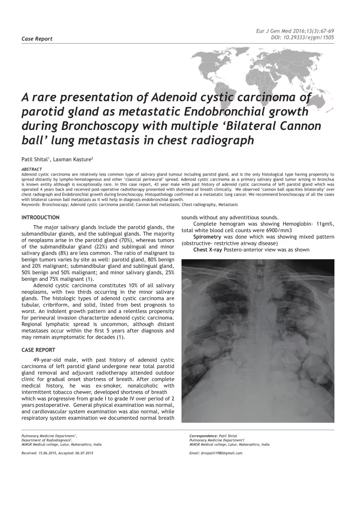
for perineural invasion characterize adenoid cystic carcinoma. Regional lymphatic spread is uncommon, although distant metastases occur within the fjrst 5 years after diagnosis and may remain asymptomatic for decades (1). CASE REPORT 49-year-old male, with past history of adenoid cystic carcinoma of left parotid gland undergone near total parotid gland removal and adjuvant radiotherapy attended outdoor clinic for gradual onset shortness of breath. After complete medical history, he was ex-smoker, nonalcoholic with intermittent tobacco chewer, developed shortness of breath which was progressive from grade I to grade IV over period of 2 years postoperative. General physical examination was normal, and cardiovascular system examination was also normal, while respiratory system examination we documented normal breath sounds without any adventitious sounds. Complete hemogram was showing Hemoglobin- 11gm%, total white blood cell counts were 6900/mm3 Spirometry was done which was showing mixed pattern (obstructive- restrictive airway disease) Chest X-ray Postero-anterior view was as shown
A rare presentation of Adenoid cystic carcinoma of parotid gland as metastatic Endobronchial growth during Bronchoscopy with multiple ‘Bilateral Cannon ball’ lung metastasis in chest radiograph
Patil Shital1, Laxman Kasture2
ABSTRACT Adenoid cystic carcinoma are relatively less common type of salivary gland tumour including parotid gland, and is the only histological type having propensity to spread distantly by lympho-hematogenous and other ‘classical perineural’ spread. Adenoid cystic carcinoma as a primary salivary gland tumor arising in bronchus is known entity although is exceptionally rare. In this case report, 43 year male with past history of adenoid cystic carcinoma of left parotid gland which was
- perated 4 years back and received post-operative radiotherapy presented with shortness of breath clinically. We observed ‘cannon ball opacities bilaterally’ over
chest radiograph and Endobronchial growth during bronchoscopy. Histopathology confirmed as a metastatic lung cancer. We recommend bronchoscopy of all the cases with bilateral cannon ball metastasis as it will help in diagnosis endobronchial growth. Keywords: Bronchoscopy; Adenoid cystic carcinoma parotid; Cannon ball metastasis; Chest radiography, Metastasis