Ali Asghar Moeinipour et al., Int. J. Bioassays , 2015, 4 (06), - - PDF document

ali asghar moeinipour et al int j bioassays 2015 4 06
SMART_READER_LITE
LIVE PREVIEW

Ali Asghar Moeinipour et al., Int. J. Bioassays , 2015, 4 (06), - - PDF document

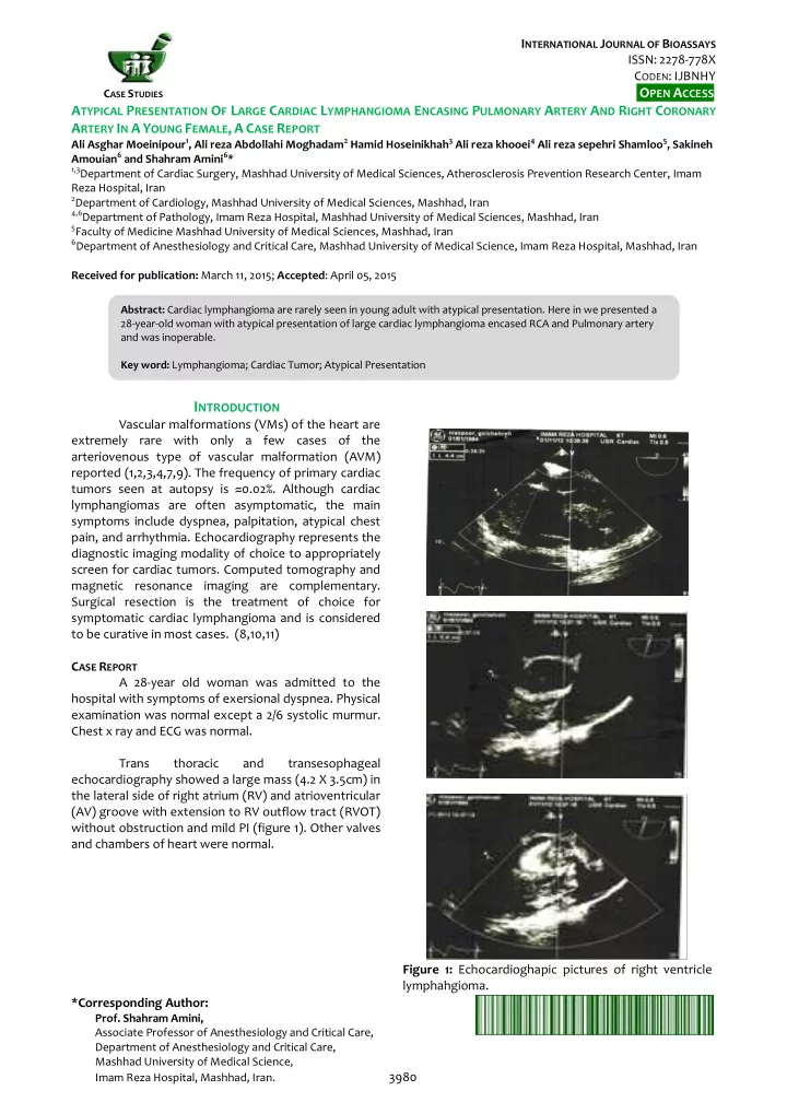
I NTERNATIONAL J OURNAL OF B IOASSAYS ISSN: 2278-778X C ODEN : IJBNHY O PEN A CCESS C ASE S TUDIES A TYPICAL P RESENTATION O F L ARGE C ARDIAC L YMPHANGIOMA E NCASING P ULMONARY A RTERY A ND R IGHT C ORONARY A RTERY I N A Y OUNG F EMALE , A C ASE R


slide-1
SLIDE 1

INTERNATIONAL JOURNAL OF BIOASSAYS ISSN: 2278-778X CODEN: IJBNHY

CASE STUDIES

OPEN ACCESS *Corresponding Author:

  • Prof. Shahram Amini,

Associate Professor of Anesthesiology and Critical Care, Department of Anesthesiology and Critical Care, Mashhad University of Medical Science, Imam Reza Hospital, Mashhad, Iran. 3980

ATYPICAL PRESENTATION OF LARGE CARDIAC LYMPHANGIOMA ENCASING PULMONARY ARTERY AND RIGHT CORONARY ARTERY IN A YOUNG FEMALE, A CASE REPORT

Ali Asghar Moeinipour1, Ali reza Abdollahi Moghadam2 Hamid Hoseinikhah3 Ali reza khooei4 Ali reza sepehri Shamloo5, Sakineh Amouian6 and Shahram Amini6*

1,3Department of Cardiac Surgery, Mashhad University of Medical Sciences, Atherosclerosis Prevention Research Center, Imam

Reza Hospital, Iran

2Department of Cardiology, Mashhad University of Medical Sciences, Mashhad, Iran 4,6Department of Pathology, Imam Reza Hospital, Mashhad University of Medical Sciences, Mashhad, Iran 5Faculty of Medicine Mashhad University of Medical Sciences, Mashhad, Iran 6Department of Anesthesiology and Critical Care, Mashhad University of Medical Science, Imam Reza Hospital, Mashhad, Iran

Received for publication: March 11, 2015; Accepted: April 05, 2015

INTRODUCTION

Vascular malformations (VMs) of the heart are extremely rare with only a few cases of the arteriovenous type of vascular malformation (AVM) reported (1,2,3,4,7,9). The frequency of primary cardiac tumors seen at autopsy is ≈0.02%. Although cardiac lymphangiomas are often asymptomatic, the main symptoms include dyspnea, palpitation, atypical chest pain, and arrhythmia. Echocardiography represents the diagnostic imaging modality of choice to appropriately screen for cardiac tumors. Computed tomography and magnetic resonance imaging are complementary. Surgical resection is the treatment of choice for symptomatic cardiac lymphangioma and is considered to be curative in most cases. (8,10,11) CASE REPORT A 28-year old woman was admitted to the hospital with symptoms of exersional dyspnea. Physical examination was normal except a 2/6 systolic murmur. Chest x ray and ECG was normal. Trans thoracic and transesophageal echocardiography showed a large mass (4.2 X 3.5cm) in the lateral side of right atrium (RV) and atrioventricular (AV) groove with extension to RV outflow tract (RVOT) without obstruction and mild PI (figure 1). Other valves and chambers of heart were normal. Figure 1: Echocardioghapic pictures of right ventricle lymphahgioma.

Abstract: Cardiac lymphangioma are rarely seen in young adult with atypical presentation. Here in we presented a 28-year-old woman with atypical presentation of large cardiac lymphangioma encased RCA and Pulmonary artery and was inoperable. Key word: Lymphangioma; Cardiac Tumor; Atypical Presentation

slide-2
SLIDE 2

Ali Asghar Moeinipour et al.,

  • Int. J. Bioassays, 2015, 4 (06), 3980-3982

www.ijbio.com 3981 Cardiac MRI showed a large intra-cavitary RV mass (85 X 43 mm) which encased the (Right coronary artery) RCA with involvement of RV anterior wall, right AV inflow, encircling RVOT with extension to pulmonic annulus and aortic root (figure 2) Figure 2: Cardiac MRI (T2) Of Right Ventricle Coronary angiography showed a normal coronary artery without vascular origin. Based on the suspicion of malignancy (angiosarcoma), cardiac biopsy was planned. After median sternotomy my finding a cluster of small vessels on the surface of RV and pulmonary valve (figure 3). Two big specimens (2*2cm) were taken and sent for frozen section and pathologic examination Frozen section showed vascular malformation without any finding of malignancy. The patient was discharged six days post operation. Figure 2: surgical view

  • f
  • f

right ventricle lymphahgioma. Pathologic examination revealed proliferation

  • f dilated vascular channels without any content with

thin wall lined by flattend endothelial cells in background of edematous fibrous stroma and focal mature lymphocytes agreggation suggestive of lymphangima (figure 4) Figure 3: Patghologic view of of right ventricle lymphahgioma. At two year follow up the patient with echocardiography was alive and with mild exersional dyspnea.

DISCUSSION

Cardiac lymphangioma is a very rare tumor of the heart, which first reported in 1911 by Armstrong and Monck berg (12). Only nine cases of cardiac lymphangioma have been reported in the medical literature (4,6,7,8). More than half of the reported cases have occurred in patients under 10 years of age (8). Mediastinal lymphangiomas are most often asymptomatic masses incidentally discovered on chest X-ray (9). The tumors most commonly occur in the pericardial space, but other unusual primary sites include the myocardium, the posterior wall of the left atrium, and the AV node regions (10). The primary site

  • f the cardiac lymphangioma in the presented case

(lower part of the interatrial septum) differs from all prior reports of this tumor (11). Cardiac lymphangioma can be successfully excised, and surgical resection is the treatment of choice for symptomatic lesions or when diagnosis is in question. The long-term outcome

  • f patients with surgically treated symptomatic lesions

is excellent. Therefore, surgical excision may be unnecessary, particularly for extensive type or asymptomatic lymphangioma.

ACKNOWLEDGMENT

We would like to thank MR. Mahmud Shariati and Hassan Rahimi and Saeed Fatehi for his contribution to preparation of this case report.

slide-3
SLIDE 3

Ali Asghar Moeinipour et al.,

  • Int. J. Bioassays, 2015, 4 (06), 3980-3982

www.ijbio.com 3982

REFERENCES

1.

Shin-Jae Kim, Eun-Seok Shin, Seon Woon Kim, Je-Kyoun Shin, Jong-Pil Cheong, Young Min Kim, andSang-Gon Lee, A Case of Cardiac Lymphangioma Presenting as a Cystic Mass in the Right Atrium Yonsei Med J 2007; 48(6): 1043

  • 1047,

2.

Danzia, Luigi Martinelli. Clinical Case Report Coronary and atrial compression by a giant cardiac hemangioma, Cardiovascular Pathology. Nucl Med Mol Imaging 2012;46(3):223–6.

3.

Aguilera B, Suárez-Mier MP, Argente T. Cardiac arteriovenous malformation causing sudden death. Cardiovasc Pathol. 2004;13:296–298.

4.

Bruce CJ. Cardiac tumors: diagnosis and management. Heart 2011;97:151–6

5.

Kemme DJ, Rainer WG. Subendocardial arteriovenous malformation in a patient with unstable angina. Clin

  • Cardiol. 1991; 14:82–84. .
  • 6. Lorine B, Meijer-Jorna, Renée BA van den Brink, Anton E

Becker, and Allard C. van der WalTwo Cases of Cardiac Arteriovenous Malformation Complicated by a Local Angioproliferative Process. Pediatr Cardiol. Aug 2010; 31(6): 868–871.

7.

Lymburner RM. Tumours of the heart: histopathological and clinical study. Can Med Assoc J. 1934;30:368–373.

  • 8. Mackie AS, Kozakewich HP, Geva T, Perez-Atayde AR,

Mulliken JB. Vascular tumors of the heart in infants and children: case series and review of the literature. Pediatr

  • Cardiol. 2005;26:344–349
  • 9. McAllister H. Tumors of the heart and pericardium. In:

Silver MD. Cardiovascular pathology. New York: Churchill Livingstone; 1983. p. 909e43

  • 10. Nataf P, Mestiri T, Martin de Lasalle E, Benomar M,

Gandjbakhch I, Cabrol C. Pericardial

  • hemolymphangioma. Apropos of a case. Arch Mal Coeur
  • Vaiss. 1988;81:1137–1140
  • 11. Nezafati MH, Javan H, Maleki MH, Hamedanchi A"

Lipomatosis of interventricular septum and both ventricles: an extremely rare pattern, Interactive cardiovascular and thoracic surgery. 12(3): 511-2

  • 12. Armstrong H, Monckeberg JG. Herzblock bedingt durch

primaren Hertztumor, bei einem funf Jahrigen Kinde. Deutsch Arch Klin Med. 1911; 102:144–166.

Source of support: Nil Conflict of interest: None Declared CITE THIS ARTICLE AS:

Ali Asghar Moeinipour, Ali reza Abdollahi Moghadam, Hamid Hoseinikhah, Ali reza khooei, Ali reza sepehri Shamloo, Sakineh Amouian and Shahram Amini, A Typical Presentation Of Large Cardiac Lymphangioma Encasing Pulmonary Artery And Right Coronary Artery In A Young Female, A Case Report, International Journal of Bioassays, 2015, 4 (06), 3980-3982.