
SLIDE 1
Page 1
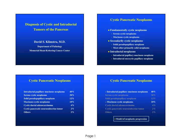
Diagnosis of Cystic and Intraductal Tumors of the Pancreas
David S. Klimstra, M.D.
Department of Pathology Memorial Sloan-Kettering Cancer Center
Cystic Pancreatic Neoplasms
Fundamentally cystic neoplasms
- Serous cystic neoplasms
- Mucinous cystic neoplasms
Secondarily cystic neoplasms
- Solid pseudopapillary neoplasm
- Most other primarily solid neoplasms
Intraductal neoplasms
- Intraductal papillary mucinous neoplasm
- Intraductal oncocytic papillary neoplasm
Cystic Pancreatic Neoplasms
Intraductal papillary mucinous neoplasms 40% Serous cystic neoplasms 30% Solid pseudopapillary neoplasm 12% Mucinous cystic neoplasms 10% Cystic ductal adenocarcinoma 4% Cystic pancreatic neuroendocrine tumor 2% Others 2%
Cystic Pancreatic Neoplasms
Intraductal papillary mucinous neoplasms
40% Serous cystic neoplasms 30% Solid pseudopapillary neoplasm 12%
Mucinous cystic neoplasms