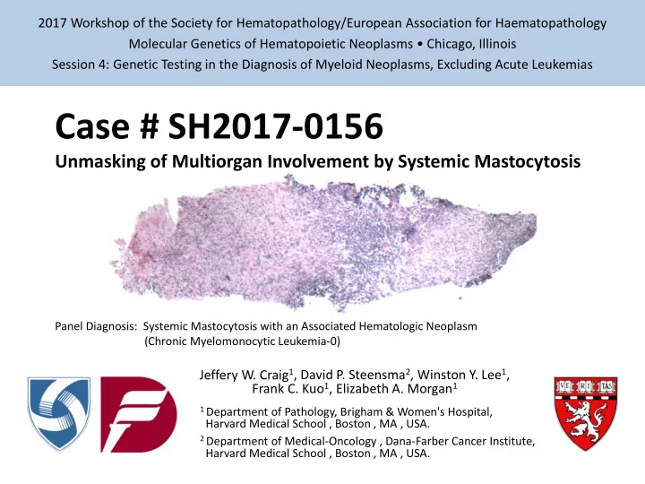

2017 Workshop of the Society for Hematopathology/European Association for Haematopathology Molecular Genetics of Hematopoietic Neoplasms • Chicago, Illinois Session 4: Genetic Testing in the Diagnosis of Myeloid Neoplasms, Excluding Acute Leukemias Case # SH2017-0156 Unmasking of Multiorgan Involvement by Systemic Mastocytosis Panel Diagnosis: Systemic Mastocytosis with an Associated Hematologic Neoplasm (Chronic Myelomonocytic Leukemia-0) Jeffery W. Craig 1 , David P. Steensma 2 , Winston Y. Lee 1 , Frank C. Kuo 1 , Elizabeth A. Morgan 1 1 Department of Pathology, Brigham & Women's Hospital, Harvard Medical School , Boston , MA , USA. 2 Department of Medical-Oncology , Dana-Farber Cancer Institute, Harvard Medical School , Boston , MA , USA.
Clinical History Patient: 72-year-old male, retired physician Presentation: LUQ pain w/ self-palpated splenomegaly, fatigue and weight loss Imaging (CT): • Splenomegaly (16.8 cm; no focal lesions) • Retroperitoneal adenopathy (≤ 2.6 cm) • Abdominal varices (c/w portal hypertension) CBC-Diff: • WBC 10.7 K/uL (H) Labs: • ALT 13 U/L • ANC 5.9 K/uL • AST 18 U/L • AMC 2.8 K/uL (H) • ALP 519 U/L (H) • HCT 35.8 % (L) • Tbili 3.9 mg/dL (H) • MCV 90.0 fL • ALB 3.3 g/dL (L) • PLT 139 K/uL (L) • PT-INR 1.5 (H) Pathology workup: • Bone marrow core biopsy • Lymph node core biopsy • Liver core biopsy Referred to DFCI for Hematology-Oncology consultation based on the BM findings: “suspicious for a myeloproliferative neoplasm”
Microscopic Findings: Bone Marrow Biopsy Preliminary Findings: • Markedly hypercellular (90%) • Myeloid hyperplasia • Megakaryocytic dysplasia • Mild reticulin fibrosis • No increase in CD34+ blasts • No ring sideroblasts Ancillary Studies: • Cytogenetics: 46,XY[20] • MDS/MPN FISH: No abnormalities
Molecular Genetic Findings Molecular analysis: Performed on peripheral blood during initial consultation “Rapid Heme Panel” (95-gene NGS assay): Results: Pathogenic Single Nucleotide Variants and Small Insertions/Deletions: • ASXL1 NM_015338 c.1926_1927insT p.G642fs* - in 70.6% of 119 reads • KIT NM_000222 c.2447A>T p.D816V - in 41.8% of 593 reads • TET2 NM_001127208 c.2596C>T p.Q866* - in 45.4% of 1442 reads • TET2 NM_001127208 c.3765C>G p.Y1255* - in 46.7% of 302 reads Concurrent CBC-Diff: • WBC 6.7 K/uL • ANC 2.4 K/uL • AMC 2.5 K/uL (H) • HCT 35.7 % (L) • MCV 95.5 fL • PLT 138 K/uL (L)
Microscopic Findings: Bone Marrow Biopsy Deeper Levels: • Scattered fibrotic foci containing intermediate-sized cells with elongated nuclei, condensed chromatin, indistinct nucleoli and abundant pale cytoplasm with well-defined cell borders
Microscopic Findings: Bone Marrow Clot Preparation Deeper Levels: • Large lymphoid aggregate surrounded by intermediate-sized cells with elongated nuclei, condensed chromatin, indistinct nucleoli and abundant pale cytoplasm with well-defined cell borders Need H&E from clot prep!
Immunohistochemistry: Bone Marrow Biopsy/Clot Prep H&E Mast cell tryptase H&E Mast cell tryptase KIT CD25 KIT CD25
Lymph Node Core Biopsy H&E Mast cell tryptase Requested for additional evaluation of unexplained lymphadenopathy following bone marrow workup Outside Interpretation: Flow cytometry: no abnormal T-cells, polyclonal B-cells Histology: No evidence of lymphoproliferative disorder KIT CD25 or malignancy “Area of irregular fibrosis with bland spindle cells consistent with myofibroblasts”; “could be reactive in nature” Post-molecular interpretation: SYSTEMIC MASTOCYTOSIS
Liver Core Biopsy H&E Mast cell tryptase Requested for additional evaluation of unexplained cholestatic liver injury and portal hypertension w/ varices & progressive ascites Hepatology workup: Negative serological evaluation (ANA, LKM, AMA, A1AT, IgG4, HepC, etc.) Outside Interpretation: Chronic biliary tract disease with prominent portal fibrous expansion, patchy mononuclear cell infiltrates and ductular reaction KIT CD25 No granulomas, bile duct inflammation or concentric periductular fibrosis Differential: primary biliary cirrhosis vs. sclerosing cholangitis (2° to CMML?) Post-molecular interpretation: SYSTEMIC MASTOCYTOSIS
Clinical Follow-Up Patient: 72-year-old male, retired physician Unifying Diagnosis: Systemic mastocytosis with an associated hematologic neoplasm (SM-AHN) • SM component: Aggressive systemic mastocytosis (ASM) • AHN component: Chronic myelomonocytic leukemia (CMML-0) Serum tryptase level elevated at 88 ng/mL (ref. <11.5 ng/mL) Treatment: Midostaurin: • Multi-kinase inhibitor capable of inhibiting KIT D816V • Effective in patients with advanced systemic mastocytosis • Initiated October 2016 Plan to delay CMML-directed therapy for as long as possible Outcome: “Continuing to get better on a regular basis” per recent clinic note • Reduction in ascites and organomegaly • Improved appetite with intentional weight gain • Increased physical activity level
Diagnostic criteria for Systemic Mastocytosis (2008 WHO Classification of tumors of hematopoietic and lymphoid tissues) Requires the major criterion and 1 minor criterion OR ≥ 3 minor criteria Major criterion: Multifocal, dense infiltrates of mast cells (≥15 mast cells in aggregates) detected in sections of bone marrow and/or other extracutaneous organ(s). Minor criteria: 1. In biopsy sections of bone marrow or other extracutaneous organs, >25% of the mast cells in the infiltrate are spindle-shaped or have atypical morphology or, of all mast cells in bone marrow aspirate smears, >25% are immature or atypical mast cells. 2. Detection of an activating point mutation at codon 816 of KIT in bone marrow, blood or another extracutaneous organ. 3. Mast cells in bone marrow, blood or other extracutaneous organs express CD2 and/or CD25 in addition to normal mast cell markers. 4. Serum total tryptase persistently exceeds 20 ng/mL (unless there is an associated clonal myeloid disorder, in which case this parameter is not valid).
Systemic Mastocytosis with an Associated Hematologic Neoplasm (SM-AHN) Diagnosis: Requires clear morphologic evidence of : (1) Systemic mastocytosis (not pure cutaneous mastocytosis) (2) An associated clonal hematologic non-MC lineage disease • Associated hematologic neoplasms (AHN) include: - Common: MDS, AML, MPN, MDS/MPN (typically CMML) - Rare: NHL, PCN Morphology is heterogeneous and largely dependent on the type of AHN May be difficult to establish in specimens where one component predominates SM may be identified retrospectively following therapy for the AHN 2 nd most common subtype of SM (after indolent systemic mastocytosis) True incidence is likely underestimated
Systemic Mastocytosis with an Associated Hematologic Neoplasm (SM-AHN) Clinical: Presentation and course generally dominated by the AHN Major exception is aggressive systemic mastocytosis, characterized by high disease burden with organomegaly and organ dysfunction Genetics: KIT mutations, especially KIT D816V, present within the MC component of the majority of SM-AHN KIT mutations variably present in the non-mast cell component of SM-AHN (CMML > AML > MPN > LPD) KIT D816V thought to function as a “differentiation inducer” or “phenotype modulator”, rather than as a strong oncogenic driver Differential Diagnosis: Non-mast cell myelogenous tumors with signs of mast cell differentiation • Tryptase-positive AML • Myelomastocytic leukemia
KIT mutations in systemic mastocytosis & other hematopoietic malignancies Systemic mastocytosis (all subtypes): >90% of cases possess gain-of-function mutations in the KIT proto-oncogene • Result in stem cell factor-independent activation of KIT • Vast majority are somatic • Rare germline mutations associated with familial mastocytosis • Majority cluster in exons 11 and 17 • Mutations in exons 8, 9, and 10 encountered infrequently Hallmark D816V mutation is seen in >80% of cases • Affects the second intracellular tyrosine kinase domain (exon 17) • D816V is resistant to imatinib • Responsive to other kinase inhibitors (e.g. midostaurin) Postulated cell of origin is a pluripotent CD34+ hematopoietic progenitor cell • KIT mutations may be confined to mast cells or present within additional hematopoietic lineages
KIT mutations in systemic mastocytosis & other hematopoietic malignancies Other hematopoietic malignancies: KIT mutations are also seen in AML with t(8;21) or inv(16) • 20% of core-binding factor AML • Impart poor prognosis KIT mutations are rarely reported in other myeloid malignancies (<5%) • e.g. MDS, MPN, other acute leukemias • Often viewed as a marker of molecular progression • Unknown how frequently such mutations represent undetected involvement by systemic mastocytosis
Recommend
More recommend