What is ultrasound? piezo-electric effect Ultrasound is energy! a - - PowerPoint PPT Presentation

what is ultrasound
SMART_READER_LITE
LIVE PREVIEW

What is ultrasound? piezo-electric effect Ultrasound is energy! a - - PowerPoint PPT Presentation

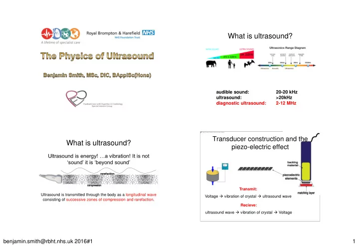
What is ultrasound? audible sound: 20-20 kHz ultrasound: >20kHz diagnostic ultrasound: 2-12 MHz Transducer construction and the What is ultrasound? piezo-electric effect Ultrasound is energy! a vibration! It is not sound it is


slide-1
SLIDE 1

benjamin.smith@rbht.nhs.uk 2016#1 1

What is ultrasound?

audible sound: 20-20 kHz ultrasound: >20kHz diagnostic ultrasound: 2-12 MHz

What is ultrasound?

Ultrasound is energy! …a vibration! It is not ‘sound’ it is ‘beyond sound’

Ultrasound is transmitted through the body as a longitudinal wave consisting of successive zones of compression and rarefaction.

Transducer construction and the piezo-electric effect

Transmit: Voltage  vibration of crystal  ultrasound wave Recieve: ultrasound wave  vibration of crystal  Voltage

slide-2
SLIDE 2

benjamin.smith@rbht.nhs.uk 2016#1 2

Transducer construction and the piezo-electric effect

Beam focussing: Sequential innervation of the outermost elements to the innermost

Transducer construction and the piezo-electric effect

Beam steering: Sequential innervation from

  • ne side to the other

Matrix Array Transducers

Transducer construction and the piezo-electric effect

matching layer

thin layer between the piezoelectric elements and the skin “accoustic matching” (we will talk briefly about this tomorrow…) reduces reflection  less attenuation and more energy transmitted

slide-3
SLIDE 3

benjamin.smith@rbht.nhs.uk 2016#1 3

Transducer construction and the piezo-electric effect

Backing material

Reduces/damps “ringing” of the piezoelectric element and thereby shortens the pulse duration  improves axial resolution. However, this comes at the expense of increasing the bandwidth.

Ultrasound frequency transmission

0 1 2 3 4 5 6 7 8 0 1 2 3 4 5 6 7 8 0 1 2 3 4 5 6 7 8 0 1 2 3 4 5 6 7 8 0 1 2 3 4 5 6 7 8 0 1 2 3 4 5 6 7 8

Shorter pulse Broad bandwidth Longer pulse Narrow bandwidth

Depth discrimination

Assumption: the speed of sound (c) in tissue is a constant 1540m/s. So that we can calculate distance to a reflection by the time elapsed.

air 330m/s fat 1480m/s soft tissue (average) 1540m/s blood 1575m/s bone 4080m/s The speed of sound is determined by the compressibility and density of that medium.

depth = ct 2 depth = ct 2 Depth discrimination

Time

20µs 40µs 60µs

Depth?

slide-4
SLIDE 4

benjamin.smith@rbht.nhs.uk 2016#1 4

The ‘modes’

A-mode – Amplitude mode B-mode – Brightness mode M-Mode – Motion mode

Temporal resolution

…. is the ability of the ultrasound machine to accurately determine the position of a moving reflector at a particular time = FRAME RATE

Temporal resolution: frames and frame rate

The pulse repetition frequency (PRF) is the number of pulses emitted per second and is dictated by depth so FR is limited by depth.

PRFmax = c 2D

Temporal resolution: frames and frame rate

A frame consists of an accumulation of pulses/scan lines. FR is limited by line density and sector width.

slide-5
SLIDE 5

benjamin.smith@rbht.nhs.uk 2016#1 5

London 2012 Womens Triathlon 1.5km swim, 40km cycle and 10km run Who won? (a) Nicola Sprig of Switzerland (top in black) (b) Lisa Nordén of Sweden (closer in blue) (c) It was a dead heat

So…. What ‘mode’ has the best temporal resolution?

m-mode

Bonus question: what is the line

density of m-mode? Can you change frame rate on your ultrasound machine?

PRFmax = c 2D

To increase our frame rate without changing depth or width, we can

  • nly do it at the expense of line density

What if we wanted to have a higher frame rate? What do we sacrifice? So for a 10cm image, we can get 1540/(2x0.1) =7700 lines. If we want 350 lines per frame segment we get 7700/350 = 22 frames per second. If we want to double our frame rate to 44Hz: Lines per segment = 7700/44 = 175 lines/frame

line density Can you change line density on your ultrasound machine?

Res = lateral resolution i.e. line density Spd = speed i.e. frame rate

↑FR ↓line density ↓lateral resolution

slide-6
SLIDE 6

benjamin.smith@rbht.nhs.uk 2016#1 6

Zoom: Reading vs Writing…

Write zoom Read zoom

Write Zoom Read Zoom

↑screen picture size Cropped image ↓width  ↑line density  ↑lat res ↓depth  ↑PRF Likely ↑ FR ↑screen picture size Whole original image continues to be captured Pixels magnified No change in FR/lat res

Temporal resolution: frames and frame rate

FR is reduced when multifocus is in use due to multiple pulses per scan line.

Temporal vs lateral resolution

To improve frame rate you can: ↓ sector width ↓ depth use write zoom (effectively ↓ width ± ↓depth) X turn off multifocus Or, reduce line density but this will be at the expense of lateral resolution.

slide-7
SLIDE 7

benjamin.smith@rbht.nhs.uk 2016#1 7

Frame rate and parallel processing

Data acquisition rate limited by speed of sound and therefore PRF. Instead  parallel processing allows multiple lines to be acquired and therefore increases FR and/or line density. How? transmission of a less focused "fatter" beam then receiving multiple simultaneous “narrow" beams. Enables the data acquisition rate to increase through the simultaneous acquisition of B-mode image lines from each individual broadened transmit pulse.

“borrowed” from Philips nSight White Paper

Lateral resolution

…. the ability to distinguish two reflectors in the direction perpendicular to the ultrasound beam. = BEAMWIDTH

poor good lateral resolution

Transmission and lateral resolution

Assumption: all echos arise from a central ultrasound beam. Lateral resolution is related to beamwidth and is best where the beam is at its narrowest, i.e. at the point of focus. Beamwidth at the focus is narrower at higher frequencies, therefore lateral resolution is better at higher frequencies. Lateral resolution is better with increased line density, i.e. less space between scan lines. Lateral resolution is worse at greater depths and beyond the focus.

slide-8
SLIDE 8

benjamin.smith@rbht.nhs.uk 2016#1 8

Axial resolution

…. the ability to distinguish between two closely spaced reflectors along the axis (i.e. in the direction) of the ultrasound beam. Spatial pulse length = λ.n (wavelength multiplied by the number of cycles within a pulse) Axial resolution = spatial pulse length/2 = λ.n/2

Transmission: axial resolution

Axial resolution depends on the physical length of the pulse and is related to frequency

c = f λ if ↑f then ↓λ ↓ pulse length →better axial resolution ↓ pulse length →better axial resolution (unable to be manually controlled)

Spatial Pulse Length

½SPL ½SPL SPL

poor good axial resolution

How to improve axial resolution?

  • Use a higher frequency transducer
  • Utilise the higher frequency component
  • f the broadband (i.e. manually adjust

frequency range)

  • Turn off harmonics

When a high amplitude ultrasound disturbance passes through an elastic medium it travels faster during the higher density compression phase than the lower density rarefaction phase causing harmonic distortions. Progressively stronger harmonic component with distance travelled. PRO: reduction in artifacts, improved signal-to-noise ratio and slight improvement in lateral resolution. CON: reduced axial resolution due to longer initial pulse length

Harmonic Imaging

slide-9
SLIDE 9

benjamin.smith@rbht.nhs.uk 2016#1 9

Harmonic Imaging OFF Harmonic Imaging ON Harmonic Imaging OFF Harmonic Imaging ON

Transmission: grating artifacts

Assumption: all echos arise from the central axis of the ultrasound beam

This is what should have happened when you made adjustments on your ultrasound machine:

On 2D, from shallow, increase the depth. What happened to the frame rate? ↓ On 2D, increase the sector width. What happened to the frame rate? ↓ On 2D, is there a way to manually change the frame rate? (does changing frame rate in this way come at the expense of anything?) Y (line density/lateral

resolution)

On 2D, turn on multifocus. What happened to the frame rate? ↓ *There should be 2 types of zoom, see which one gives you a better image. Are you able to use one of these zoom modes after the image is captured? ‘write’zoom (the one which crops the image) *On 2D, move the focus up and down, do you notice a difference? Y (reduced lateral resolution beyond focus) *On 2D, change transducers/frequency. Which has better image strength? Low freq (we’ll talk about this tomorrow) *On 2D, change transducers/frequency. Which has better image sharpness? High freq