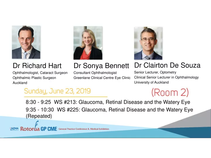
Dr Clairton De Souza Dr Richard Hart Dr Sonya Bennett Senior - - PowerPoint PPT Presentation

dr clairton de souza dr richard hart dr sonya bennett
SMART_READER_LITE
LIVE PREVIEW

Dr Clairton De Souza Dr Richard Hart Dr Sonya Bennett Senior - - PowerPoint PPT Presentation

Dr Clairton De Souza Dr Richard Hart Dr Sonya Bennett Senior Lecturer, Optometry Ophthalmologist, Cataract Surgeon Consultant Ophthalmologist Clinical Senior Lecturer in Ophthalmology Ophthalmic Plastic Surgeon Greenlane Clinical Centre Eye


slide-1
SLIDE 1

Dr Richard Hart

Ophthalmologist, Cataract Surgeon Ophthalmic Plastic Surgeon Auckland

8:30 - 9:25 WS #213: Glaucoma, Retinal Disease and the Watery Eye 9:35 - 10:30 WS #225: Glaucoma, Retinal Disease and the Watery Eye (Repeated)

Dr Sonya Bennett

Consultant Ophthalmologist Greenlane Clinical Centre Eye Clinic

Dr Clairton De Souza

Senior Lecturer, Optometry Clinical Senior Lecturer in Ophthalmology University of Auckland

slide-2
SLIDE 2

Retinal Detachment

Dr Clairton F. de Souza

slide-3
SLIDE 3

(Rhegmatogenous) Retinal Detachment

 Major retinal disorder requiring surgery

  • Annual incidence of 11.8 cases per 100.000 in northern

New Zealand (Polkinghorne and Craig 2004)  Major cause of severe loss of vision  Success rate of primary surgical repair: 85%; (95% after 2-3 surgeries)

slide-4
SLIDE 4

Idiopathic Polypoidal Choroidal Vasculopathy

slide-5
SLIDE 5

Retinal Detachment – Macula-on

slide-6
SLIDE 6
slide-7
SLIDE 7

Vision: 6/24 Vision: 6/9

slide-8
SLIDE 8
  • Dr. Didier Ducournau – Nantes, France
slide-9
SLIDE 9

Retinal Detachment

Loss of central vision

Detached Retina Attached Retina

Fovea

slide-10
SLIDE 10

Normal Vision:

6/6 Vision in RRD: Counting fingers Vision after RRD repair: 6/15

slide-11
SLIDE 11

Retinal Cell Biology Lab – Institute of Neuroscience, Santa Barbara California

slide-12
SLIDE 12

Ocular trauma and retinal detachment

Iridodialysi s Lens subluxation

slide-13
SLIDE 13

Does Bungee jumping cause retinal detachment?

  • No documented association between bungee

jumping and retinal detachment

Gravitational force (G-force) Rapid - 3G deceleration Intravenous pressure: +100mmHg

All papers report: Ocular haemorrhages (conjunctival and retinal)

Valsalva Phenomenon

slide-14
SLIDE 14

Does Bungee jumping cause retinal detachment?

  • No documented association between bungee

jumping and retinal detachment

Gravitational force (G-force) Rapid - 3G deceleration Intravenous pressure: +100mmHg

All papers report: Ocular haemorrhages (conjunctival and retinal)

Valsalva Phenomenon

slide-15
SLIDE 15

Is it preferable for a high myope pregnant to give birth by C-section? No.

High myopes and even women with history

  • f retinal break treatment do not require C-section.

Valsalva manœuvres during labour do not increase risk of retinal detachment Hormonal and Immunological changes during pregnancy do not increase risk of retinal tear/detachment in high myopic women. Is she at higher risk of retinal detachment after normal labour?

slide-16
SLIDE 16

Importance of Awareness of retinal detachment symptoms

slide-17
SLIDE 17

Floaters – Ophthalmologist view

slide-18
SLIDE 18

Flashes (Photopsia) - Differential Diagnoses

Diagnosis Duration Colour Location Shape Stimulus Other symptoms Flickering PVD Seconds

  • r less

White Temporal Periphery Crescentic vertical Eye Movements Floaters No Migraine 20-30 minutes Not typical Paracentral Arcuate, zigzag Stress, food Scotoma, headache, nausea Yes Occipital ischaemia Minutes Nil Central Petaloid Neck movements, exertion Yes Cystoid Macular Oedema Constant Variable Central Pinpricks Nil Poor vision Yes Outer retinal

  • r RPE

abnormality Minutes Blue/purple Paracentral Blob, spirals Nil Scotoma No Retinal detachment Seconds Golden Central/par acentral Comet

  • blique

horizontal Eye Movements Visual field loss No

slide-19
SLIDE 19
slide-20
SLIDE 20

Retinal detachment - Summary

  • RD is a potentially blinding condition if untreated.
  • Surgical success rates are above 80% with a single

surgery for non-complex cases (95% with 2 or more surgeries).

  • Patient awareness, fast recognition of suggestive

symptoms and prompt referral are crucial for optimal visual prognosis.

slide-21
SLIDE 21

Diagnosis Duration Colour Location Shape Stimulus Other symptoms Flickering PVD Seconds

  • r less

White Temporal Periphery Crescenti c vertical Eye Movements Floaters No Migraine 20-30 minutes Not typical Paracentral Arcuate, zigzag Stress, food Scotoma, headache, nausea Yes Occipital ischaemia Minutes Nil Central Petaloid Neck movements, exertion Yes Cystoid Macular Oedema Constant Variable Central Pinpricks Nil Poor vision Yes Outer retinal

  • r RPE

abnormality Minutes Blue/purple Paracentral Blob, spirals Nil Scotoma No Retinal detachment Seconds Golden Central/par acentral Comet

  • blique

horizontal Eye Movements Visual field loss No

Flashes (Photopsia) - Differential Diagnoses