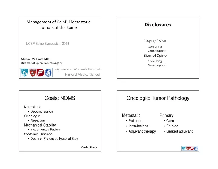

Management of Painful Metastatic Disclosures Tumors of the Spine Depuy Spine UCSF Spine Symposium 2013 Consulting Grant support Biomet Spine Michael W. Groff, MD Consulting Director of Spinal Neurosurgery Grant support Brigham and Woman’s Hospital Harvard Medical School Goals: NOMS Oncologic: Tumor Pathology Neurologic • Decompression Metastatic Primary Oncologic • Resection • Paliation • Cure Mechanical Stability • Intra-lesional • En bloc • Instrumented Fusion • Adjuvant therapy • Limited adjuvant Systemic Disease • Death or Prolonged Hospital Stay Mark Bilsky
Metastatic Cord Compression Incidence of Spinal Metastases Histology is unknown at presentation in 10% 40-60% of cancer patients harbor spinal of cases with metastatic spinal cord metastasis compression • 10% are symptomatic 50% of those cases will turn out to have lung 18,000 new cases annually in NA cancer Most common malignant spine tumor Most common site of bone metastasis Primary Tumor Spinal Level Breast 22% Thoracic 70% Lung 15% Lumbar 20% Myeloma 9% Cervical 10% Lymphoma 7% Probability of met = GI 5% f (number of bodies, Prostate 10% size of bodies) Renal 6% Thyroid 3%
Location Within Body Manifestations of Spine Metastases Back Pain Vertebral body 85% Cord Compression • Early involvement of pedicle Pathologic fracture - 8% incidence Paravertebral 15% • 53% are breast cancer Epidural space 5% Intradural rare “ Metastatic ” Back Pain Pathologic Fracture Most common presentation Pain onset to neuro signs Constant dull ache avg 7 mos Progresses to frank pain [Hatrick et al, Radiother Onc 2000] Awakens from sleep Aggravated by valsalva Etiology • Tumor spread/tissue destruction • Periosteal innervation • Cord compression • Spinal instability • Nerve root irritation 50% destruction required [Edelstyn et al, Clin Radiol 1967]
Results of Treatment for Spinal Cord Compression: Results of Treatment for Spinal Cord Compression: Pathologic Fracture Radiotherapy Alone Radiotherapy Alone Authors Year Patients (n) % Improved % Worse Mones et al. 1966 41 34 - Khan et al. 1967 82 42 - Cobb et al. 1977 18 50 22 Gilbert et al. 1977 29 41 21 Marshall & Langfitt 1977 130 49 - Greenberg et al. 1980 83 57 7 Stark et al. 1982 31 35 - Constans et al. 1983 108 39 26 Obbens et al. 1984 83 28 23 Harrison et al. 1985 33 27 36 Bach et al. 1990 149 35 18 Maranzano 1995 209 76 0 Mean - 44 17 Total 996 Results of Treatment for Spinal Cord Compression: Results of Treatment for Spinal Cord Compression: Results of Treatment for Spinal Cord Compression: Results of Treatment for Spinal Cord Compression: Laminectomy with or without Radiotherapy Laminectomy with or without Radiotherapy Laminectomy (Posterior Decompression) and Stabilization Laminectomy (Posterior Decompression) and Stabilization Authors Year Patients (n) % Improved % Mortality % Worse % Motor % Pain Hall & McKay 1973 129 30 - - Year Patients (n) Authors % Mortality Improved Improved Brady et al. 1975 90 61 - - - Brunon et al. 1975 20 100 - Merrin et al. 1976 22 22 0 0 84 100 Cobb et al. 1977 26 46 23 - Hansebout et al. 1980 82 - Gilbert et al. 1977 65 45 - - 65 100 Miles et al. 1984 23 - Marshall & Langfitt 1977 17 29 - - 45 65 DeWald et al. 1985 17 6 Gianotta & Kindt 1978 33 30 18 12 75 - Kleinman et al. 1978 20 15 5 15 Overby et al. 1985 12 - Livingston & Perrin 1978 100 58 - 0 48 - Solini et al. 1985 33 3 Baldini et al. 1979 140 30 19 0 70 79 Heller et al. 1986 33 - Gorter 1979 31 39 - 13 82 80 Perrin et al. 1987 200 8 Dunn et al. 1980 104 33 23 10 Levy et al. 1982 39 82 15 8 1996 38 100 Olerud 51 0 Stark et al. 1982 84 37 - - 1997 76 0 Bauer 67 - Constans et al. 1983 465 46 13 - Bilsky 1999 25 90 - 12 Klein et al. 1984 194 54 16 - Kollmann et al. 1984 103 56 - - Mean 67 89 8 Garcia-Picazo et al. 1990 53 41 - - Back et al. 1990 91 59 11 - Total 563 Landmann et al. 1992 127 58 2 Mean 44 13 7 Total 1933
Results of Treatment for Spinal Cord Compression: Results of Treatment for Spinal Cord Compression: Management Goals Vertebral Body Resection and Stabilization Vertebral Body Resection and Stabilization Improved Improved % Authors Year Patients (n) Motor % Pain % Mortality 7 Slatkin and Posner 1982 29 56 60 Neurologic function 6 Harrington 1984 52 65 80 Siegal and Siegal 1985 61 80 91 6 Oncologic Sundaresan et al. 1985 101 70 85 8 Onimus et al. 1986 36 72 97 6 • No effect on longevity Perrin & McBroom 1987 21 95 90 5 Mechanical stability Moore & Uttley 1989 26 62 71 30 Sundaresan et al. 1991 54 100 90 6 • Intractable pain Hall & Webb 1991 15 86 - 20 • Risk of collapse Fidler 1994 18 93 94 20 Hosono et al. 1995 90 81 94 0 (Systemic disease) Gokaslan et al. 1998 72 78 92 3 Mean 76 85 10 Total 575 Work-up Localization History Root C5 C6 C7 L4 L5 S1 • Ambulation, B/B, Pain worse at night Physical exam • LE weakness, long tract signs, sensory level, Motor Dorsiflexion Deltoid Biceps Triceps Quads Plantarflexion point tenderness EHL MRI of the spine - compression CT of the spine - stability Thumb & Lat leg, Upper 3ed Anterior Restage primary Sensory index dorsum of Sole of foot arm digit thigh • CT chest, abdomen, and pelvis finger foot Labs with LFT ’ s • FDG PET • 10 - 30% with synchronous lesions Reflex Biceps Supinator Triceps Patelar None Achillies
Case EW Stability Assessment Anatomic • 2 column 66 year old man • 3 Column C2 metastasis of non- Physiologic small cell lung CA • History positional pain Severe neck pain Trauma is not Oncology Neurologically intact Metastasis to liver Harms What to do? Do No Harm
Case CM CM intraop 48 year old woman LE weakness, back pain Metastatic osteosarcoma from hip What to do? Post op - 1 year MRI Study of choice Unsuspected lesions in 30% Whole body fast-imaging techniques: • STIR 30-45 min • Echo-planar 6 min False positives
Radionuclide Imaging 55 yo prostate CA & LBP 50-80% more sensitive than plain films Detection up to 18 mos earlier [Pagani & Libshitz, Radiol Clin North Am 1982] 2% cold SPECT MRI vs Bone Scan Bone scan assesses cortical bone SPECT more sensitive, Early lesions small and intramedullary better localization MRI more Benign: ↑ uptake in sensitive endplate, lateral body border, facet, spinous Bone scans process more cost Malignant: ↑ uptake in effective pedicle, central body, entire vertebra, cold lesions with ↑ uptake at margins Taoka et al, AJR 2001 [Sedonja et al, Clin Nucl Med 1999]
Myelography Importance of Biopsy Metabolic bone disease Risk of LP below hi- (osteoporosis & osteomalacia) grade block Benign tumors Use high cervical Myeloma/plasmacytoma cisternal puncture Paget ’ s disease Primary bone sarcomas Be prepared for emergent OR DDX - CT guided biopsy • Core bx preferable to FNA Breast CA Intraop - LECA 50 year old woman Back and leg pain Worse s/p XRT Known Breast CA Only known met
IntraOp Postop Renal Cell CA <10% cases Median survival 6-9 months historically 5-yr survival 10-50% 10-yr survival 5-30% Improved adjuvant therapy Standard radiation therapy not effective Conformal radiation Preop embolization very helpful
Renal Cell CT Myelogram 59 year old woman S/P T12 resection of renal cell met Index resection shortly after nephrectomy Local recurrence 3 years post op Presents w/ paraparesis, kyphosis, HW failure Postop Immediately postop worse RLE weakness Ambulating without assistance at 3 months Importance of embolization • Prabhu et al JNS 2003 • Jackson et al JNS 2001
Study Design All patients • Underwent MRI • Treated with Decadron • Diagnosis confirmed with biopsy Stratification based on • Tumor type • Ambulatory status • Spine stability Randomized to • surgery with XRT or XRT alone (30Gy) Treatment Entry Criteria Radiation started within 24 hours Known cancer Surgery within 24 hours Symptomatic lesion Goals Resectable based on MRI • Remove as much tumor as possible Not paraplegic > 48 hours • Immediate decompression No prior XRT • Stabilization when needed Exclusion: lymphoma, leukemia, multiple XRT started less than 14 days post-op myeloma, germ cell tumor, primary spinal tumor
Endpoints Results Ambulation Primary • Surgery + XRT 126 days • Ambulation • XRT alone 35 days • For non-ambulatory patients 56% vs 19% Secondary • Continent Continent • Surgery + XRT 142 days • ASIA and Frankel grade • XRT alone 12 days • Narcotic and steroid use Survival • Survival • Surgery + XRT 129 days • XRT alone 100 days Results Conclusion Steroid use 1.6mgs vs 4.2 mgs Surgery + XRT is superior to XRT alone for Narcotic use 0.4 mgs Morphene vs 4.8 mgs symptomatic Spinal Column Metastasis. Complications Post-op 12% • Hardware/ fusion - half • Wound breakdown – half
Vertebroplasty for Tumor VP/KP Originally applied to osteoporotic burst fractures Addresses the structural deficiencies of the anterior column An adjunct to either • XRT • Surgery Complications Anterior Column Augmentation T4
Recommend
More recommend