
SLIDE 1
1
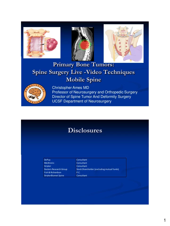
Primary Bone Tumors: Spine Surgery Live -Video Techniques Mobile Spine
Christopher Ames MD Professor of Neurosurgery and Orthopedic Surgery Director of Spine Tumor And Deformity Surgery UCSF Department of Neurosurgery
Disclosures
DePuy Medtronic Stryker Docters Research Group Fish & Richardson StrykerBiomet Spine Consultant Consultant Consultant Stock Shareholder (excluding mutual funds) P.C. Consultant