
SLIDE 1
9/14/2019 1
Arrhythmogenic Right Ventricular Cardiomyopathy: Diagnosis
Harikrishna Tandri Associate Professor of Medicine Director of VT Ablation Johns Hopkins Medical Institutions
www.ARVD.com
Disclosures
- Abbott Labs: Grant support
Objectives
- Provide overview of ARVC clinical
presentation and diagnosis
- Describe the role of task force criteria
- Describe MRI features of ARVC
- Role of electroanatomic mapping
- Concept of Arrhythmogenic
Cardiomyopathy
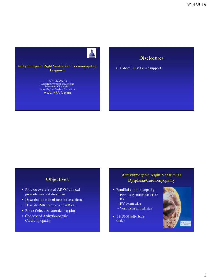
- Familial cardiomyopathy
– Fibro-fatty infiltration of the RV – RV dysfunction – Ventricular arrhythmias
Arrhythmogenic Right Ventricular Dysplasia/Cardiomyopathy
- 1 in 5000 individuals