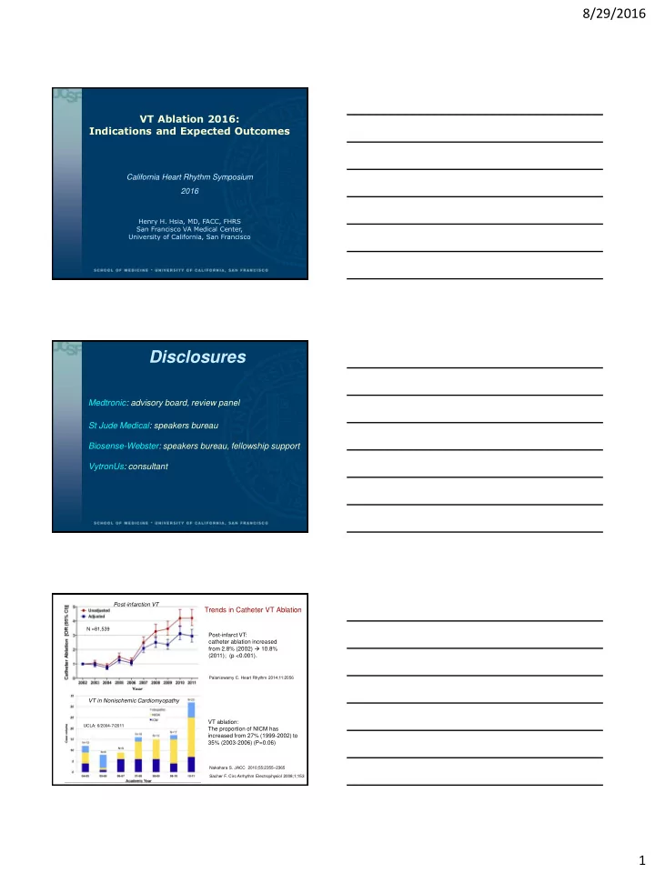
disclosures
play

Disclosures Medtronic: advisory board, review panel St Jude - PDF document

8/29/2016 VT Ablation 2016: Indications and Expected Outcomes California Heart Rhythm Symposium 2016 Henry H. Hsia, MD, FACC, FHRS San Francisco VA Medical Center, University of California, San Francisco Disclosures Medtronic: advisory


  1. 8/29/2016 VT Ablation 2016: Indications and Expected Outcomes California Heart Rhythm Symposium 2016 Henry H. Hsia, MD, FACC, FHRS San Francisco VA Medical Center, University of California, San Francisco Disclosures Medtronic: advisory board, review panel St Jude Medical: speakers bureau Biosense-Webster: speakers bureau, fellowship support VytronUs: consultant Post-infarction VT Trends in Catheter VT Ablation N =81,539 Post-infarct VT: catheter ablation increased from 2.8% (2002)  10.8% (2011); (p <0.001). Palaniswamy C. Heart Rhythm 2014;11:2056 VT in Nonischemic Cardiomyopathy VT ablation: UCLA: 6/2004-7/2011 The proportion of NICM has increased from 27% (1999-2002) to 35% (2003-2006) (P=0.06) Nakahara S. JACC 2010;55:2355 – 2365 Sacher F. Circ Arrhythm Electrophysiol 2008;1;153 1

  2. 8/29/2016 Anatomical Substrate Post-Myocardial Slow conduction in the infarcted tissue, Infarction with ‘zigzag' course of activation Tung R. Circulation 2011;123:2284 deBakker J. Circulation 1988; 77:589 deBakker J. Circulation 1993;88;915 RBRS VT: Entrainment with Concealed Fusion  Isthmus, #196 Sti-QRS=Egm-QRS 142 msec I II III AVR AVL AVF sti-QRS Egm-QRS 142 ms 142ms V 1 V 2 V 3 V 4 V 5 V 6 Abl d PPI=360 ms VTCL=363 ms Abl p His d His p RV Inferolateral Scar AoV RBRS VT Ent 170 Outer 255 Loop LP 180ms Exit 190 198 Isth Dysynchrony on ICE 2

  3. 8/29/2016 Ablation at Isthmus: RBRS VT Termination in 1.5 sec I II III AVR AVL AVF V 1 V 2 V 3 V 4 V 5 V 6 Abl d Abl p His d His p RV Stim 0.5-1.6 mV 0.36-0.55 mV 0.21-0.31 mV I II III AVR AVL AVF V 1 Late potentials with V 2 V 3 decremental local V 4 conduction delay V 5 V 6 Abl d Abl p His RVA I II III aVR aVL Pacemap aVF Spontaneous within the V 1 LBB-RI VT channel V 2 V 3 V 4 V 5 V 6 LP 180 ms LP 171 ms LP 194 ms Decremental LP delay Perfect pacemap 0.21-0.31 mV 0.5-1.6 mV 3

  4. 8/29/2016 Kaplan – Meier Estimate of Survival Free from Kaplan-Meier Estimates for Survival ICD (Shock & ATP) Therapy Free from VT or VF Ablation P=0.045 Control P=0.007 Ablation Control SMASH VT Trial VTACH Trial Thermocool VT Ablation Trial N=231, VT (median, 11 in preceding 6 mo), primary end point of freedom from VT after 6 month f/u. VT episodes were reduced from a median of 11.5 to 0 (P<0.0001). Reddy VY. NEJM 2007;357:2657-65. Kuck KH. Lancet 2010; 375: 31 – 40 Stevenson WG. Circ 2008;118:2773-82 Catheter Ablation in Post-infarct VT LVEF Acute Follow-up Recurrent Adverse N Indications (%) success (months) VT/ICD Rx events SMASH-VT Recur/induc 64 31±10 ----- 24 13%* 4.6% (2007) VT/VF ThermoCool 231 MMVT 25 49% 6 47% 7.3% (2008) VTACH 52 Stable VT 34±9.6 ----- 21.9±8.3 53% 3.8% (2010) Euro-VT 63 Recur VT 30±13 81% 12±3 49% 5% (2010) Yokokawa Recur VT 98 27±13 63% 35±23 34% 7.1% (2012) ICD Rx Silberbauer Drug 155 31±9.4 ----- ~19 32% 7% refractory VT (2014) Dinov 164 Recur VT 32±11 77.4% 27 43% 11.1% (2014) VT Recurrences After the Ablation Procedure Thermocool VT Ablation (2008) Yokokawa et al (2012) Success Failure No recurr Recurrent p p (123) (108) VT (65) VT (33) 65 69 LVEF (%) 29 ± 14 25 ± 12 0.26 Age 0.012 (58-70) (62-73) Anterior MI 18 (28%) 14 (42%) 0.14 Heart failure 52% 73% 0.002 Scar area 25 25 69 ± 30 93 ± 40 0.002 LVEF (%) 0.387 (cm 3 ) (20-35) (15-35) # clinical VT 4 ± 5 3 ± 3 0.29 Multiple MI 5% 14% 0.016 Clinical VT VT events in 10 14 359 ± 73 350 ± 77 0.34 0.37 prec 6 mo (4-30) (6-38) CL # induced 3 4 # induced 13 (20%) 9 (27%) 0.41 0.002 VT/pt (2-4) (3-6) VT Longest VT 440 450 Identified 3 ± 2 4 ± 3 0.49 0.251 CL (370-500) (380-538) critical sites Shortest VT 330 305 RF duration 63 ± 44 73 ± 48 0.35 0.029 CL (271-400) (272-350) (min) Total # RF 24 26 Postop VT- 24/63 (38%) 11/32 (34%) 0.72 0.029 nonclinical lesions (11-32) (16-39) Postop VT Postop VT- 0/63 (0%) 0/63 (0%) 1.0 30 58 <0.001 clinical Induction 4

  5. 8/29/2016 VT Recurrence Rate vs Ablation Strategies Recurrence Rate During Follow-up 13.4 ± 4 months Complete LP abolition 100 No VT inducible All Patients (n=50) 90 Incomplete LP abolition Percentage of Patients (%) 75% 80 Persistent VT inducibility 70 60 50% 50 40 30 20 12.5% 9.5% 10 0 Silberbauer J et al. Circ Arrhythm Electrophysiol, 2014. 7(3):424-435 Local Abnormal Ventricular Activities 1 2 3 4 (LAVA) 5 Regional Variation of LAVA Latency Freedom from Recurrent VT or Death * Latency of LAVA is affected by locations. Only 3% of septal LAVA were separated from far-field ventricular egm Jais P. Circulation 2012;125:2184 Komatsu Y. Heart Rhythm 2013;10:1630 Ventricular Arrhythmia/ICD Therapy-Free Survival by the Ablation Approach In Post-infarct VT N=92, f/u of 25 ± 10 months N= 160, f/u ~19 months Homogenization (Endo ± Epi): 19% 16% P<0.001 VT non-inducible+LP abolition p=0.006 Endo substrate ablation: 47% VT non-inducible, no LP abolition VT inducible DiBiase L . JACC 2012;60:132 Silberbauer J. Circ Arrhythm Electrophysiol. 2014;7:424 5

  6. 8/29/2016 16.4% Scar De-channeling Berruezo A. Circ Arrhythm Electrophysiol . 2015;8:326-336 Recurrence after Catheter Ablation of Post-infarct VT Limited substrate ablation Extensive 38% RRR substrate modification Santangeli et al, Indication for Ablation and Trials, Ventricular Tachycardia Ablation: A Practical Guide, 2014. CardioText Differences Between NICM and ICM Substrates Endo Scar and Endo DS area : ICM >> NICM Endo and Epi LP : ICM >>NICM. LP-targeted ablation was more effective in ICM (82% non-recurrence at 12±10 mon f/u) vs NICM patients with less favorable outcomes (50% at 15±13 mon f/u). 101±55 4.3% 4.1% 56±33 55±41 53±28 1.3% 2.1% Nakahara S. JACC 2010;55:2355 – 2365 6

  7. 8/29/2016 Epicardial VT Ablation: A Multicenter Safety Study Characteristics of the Patient Population Ischemic Dilated Other Total ARVC No CMP CMP CMP CMP patients (n=14) (n=17) (n=51) (n=39) (n=13) (n=134) Relative to a control 16% 35% 41% 6% 18% 19% population(n=722) 48 32 9 10 10 109 Sex (Male %) (94%) (82%) (64%) (59%) (77%) (81%) 46 33 9 15 12 115 Prior endocardial ablation (90%) (85%) (64%) (88%) (92%) (86%) Epicardial mapping 42 36 14 12 9 113 and ablation (82%) (92%) (100%) (71%) (69%) (84%) Sacher F. JACC 2010; 55: 2366 Endo Scar area (cm 2 ) Epicardial vs Endocardial Low Epi Voltage Scar Distributions in Non-Ischemic Cardiomyopathy Wide/split/late egm: epicardial (49.7%) controls (2.3%). Soejima K. JACC 2004; 43;10:1834 Cano O. JACC 2009;54:799 – 808 Endo-Epicardial Mapping in Patient with Nonischemic Cardiomyopathy LV endocardium: LV epicardium: minimal scar extensive scar MV MV I II III AVR AVL AVF Entrainment with V 1 concealed fusion: V 2 Isthmus V 3 V 4 V 5 V 6 Abl d Abl p RV 7

  8. 8/29/2016 Scar Patterns and Ablation in Nonischemic Cardiomyopathy Basal anteroseptal scar (42%): -aortic root ± anteroseptal endo LV (89%) -anterior cardiac vein (11%), with -RV septum (22%) -epicardium (11%) Green: good pacemap Yellow: ECF White: VT termination Inferolateral scar (47%): -epicardium (63%) -inferolateral endo LV (37%) Piers, S. Circ Arrhythm Electrophysiol. 2013;6:875 Nonischemic Cardiomyopathy: Anteroseptal vs Inferolateral Scar Endocardial unipolar voltage: - Anteroseptal (AS): 44/87 (51%) - Inferolateral (IL): 43/87 (49%) -AS has more extensive endo unipolar scar, freq involves an intramural septal substrate. Epi LPs: common in the IL (81%) vs INFEROLATERAL GROUP AS (4%), p<0.001) and correlated with VT termination sites (p=0.014). VT recurred in 44 patients (51%) during a median f/u 1.5 years. AS scar was associated with higher VT recurrence (74% vs 25%, p<0.001) and redo procedure rates (59% vs 7%, p<0.001). Oloriz, T. Circ Arrhythm Electrophysiol. 2014;7:414-423 Outcome of Catheter Ablation in Nonischemic Cardiomyopathy F/U Acute N Approach No Recurrence Noninducibility (months) Marchlinski 8 10 Endo 1 (12%) 3 (38%) (2000) Hsia 19 22 Endo 8 (42%) 5 (26%) (2003) Soejima Endo (22) 12/22 (55%) 12/22 (55%) 22 11±9 (2004) ± Epi (7) 6/6 (100%) 4/6 (67%) Cano 14/21 (67%) 15/21 (71%) 22 18±7 Endo+Epi (22) (2009) 12/17 (71%) 12/14 (86%) Kuhne Endo (24) 67% if LP+ 24 18 45/67 (67%) (2010) ± Epi (7) 7% if LP- Della Bella Endo+Epi (57) 67 17±18 45/67 (67%) 60.7% (2011) Epi only (10) Tung Endo only (35) 40% 33% 56 12 (2013) Endo+Epi (29) 45% 36% Dinov Endo only (43) 63 20 42 (66.7%) 23% (2014) Endo+Epi (20) 8

Download Presentation
Download Policy: The content available on the website is offered to you 'AS IS' for your personal information and use only. It cannot be commercialized, licensed, or distributed on other websites without prior consent from the author. To download a presentation, simply click this link. If you encounter any difficulties during the download process, it's possible that the publisher has removed the file from their server.

Recommend


More recommend