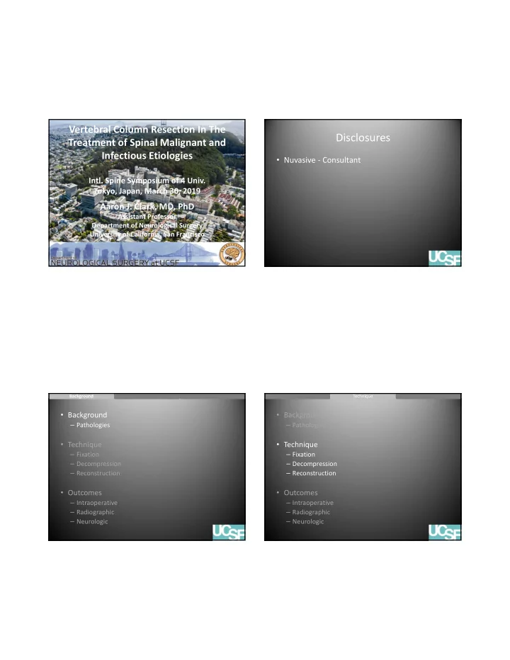

Vertebral Column Resection In The Disclosures Treatment of Spinal Malignant and Infectious Etiologies • Nuvasive ‐ Consultant Intl. Spine Symposium of 4 Univ. Tokyo, Japan, March 30, 2019 Aaron J. Clark, MD, PhD Assistant Professor Department of Neurological Surgery University of California, San Francisco Background Background Technique Technique Outcomes Outcomes Background Background Technique Technique Outcomes Outcomes • Background • Background – Pathologies – Pathologies • Technique • Technique – Fixation – Fixation – Decompression – Decompression – Reconstruction – Reconstruction • Outcomes • Outcomes – Intraoperative – Intraoperative – Radiographic – Radiographic – Neurologic – Neurologic
Background Background Technique Technique Outcomes Outcomes Background Background Technique Technique Outcomes Outcomes Background • Background – Pathologies • Pathologic fractures can be a source of pain and disability • Technique – Pain is the most common presenting symptom for – Fixation spine osteomyelitis and spinal metastatic disease – Decompression – Neurologic deficit can result from spinal cord – Reconstruction compression • Outcomes • Pathologic fractures may result in spinal – Intraoperative deformity requiring anterior column – Radiographic reconstruction – Neurologic Background Background Technique Technique Outcomes Outcomes Background Background Technique Technique Outcomes Outcomes Surgical goals Anterior reconstruction • Spinal cord decompression • Removal of pathologic tissue • Arthrodesis • Maintenance/improve ment in sagittal alignment
Background Background Technique Technique Outcomes Outcomes Background Background Technique Technique Outcomes Outcomes Background Background Technique Technique Outcomes Outcomes Background Background Technique Technique Outcomes Outcomes Thoracic corpectomy • Risks • Can a large rectangular end plate expandable cage be safely placed from an all posterior – Lung injury approach? – Vascular injury – Pneumothorax
Background Background Technique Technique Outcomes Outcomes Background Background Technique Technique Outcomes Outcomes Instrumentation levels • Resection – Circumferential bony removal – Levels dictated by spinal cord compression, bony destruction, and/or deformity • Fixation – Increase levels above and below as number of levels resected increases • 2 ‐ 4 above and below – Do not terminate at apex of thoracic kyphosis Safaee et al., 2019, Operative Neurosurgery (in press) Background Background Technique Technique Outcomes Outcomes Background Background Technique Technique Outcomes Outcomes The use of titanium cages for Aryan et al., 2007 osteomyelitis at UCSF • 15 patients with osteomyelitis (retrospective) • Corpectomy – One level (1 patient) – Two level (13 patients) – Six level (1 patient) • Follow up 11 ‐ 33 months – 0 recurrent infections – 100% fusion rate
Background Background Technique Technique Outcomes Outcomes Background Background Technique Technique Outcomes Outcomes The use of titanium cages for Lu et al., 2009 osteomyelitis at UCSF • 36 patients with osteomyelitis (retrospective) • Follow ‐ up (10 ‐ 39 months) • 2/36 (5.6%) recurrence Background Background Technique Technique Outcomes Outcomes Background Background Technique Technique Outcomes Outcomes Fixation techniques Exposure • Wide exposure • Pedicle screws – Expose transverse processes at all • Proximal junctional kyphosis prevention instrumented levels – Transverse process hooks at the cephalad level • Expose rib heads at – Posterior ligamentous complex augmentation levels of planned corpectomy
Background Background Technique Technique Outcomes Outcomes Background Background Technique Technique Outcomes Outcomes Temporary stablization Decompression • Stabilize with pedicle • Laminectomy screw based racks – Involved vertebra – Partial above and below • Allows controlled distraction and compression Background Background Technique Technique Outcomes Outcomes Background Background Technique Technique Outcomes Outcomes Decompression Rhizotomy • Bilateral facetectomy • Bilateral if possible • Rib head removal bilaterally • For T8 ‐ 12, temporary • Bilateral pediculectomy clip for 10 minutes – Usually at least partially – Check MEPs every destroyed minute
Background Background Technique Technique Outcomes Outcomes Background Background Technique Technique Outcomes Outcomes Rhizotomy Rhizotomy • For 3 level corpectomies – Divide 3 nerve roots on one side – Divide only one on the other Background Background Technique Technique Outcomes Outcomes Background Background Technique Technique Outcomes Outcomes Corpectomy Anterior reconstruction • Rectangular footprint • Define interval between the PLL and the expandable titanium thecal sac cage • Annulotomy • Remove disc and vertebral body with • Deformity correction osteotomes – 3 ‐ 5 degrees of kyphotic – Be careful to not violate the endplates angulation • Once a defect is created, remove the central • Spinal shortening region with the impactors
Background Background Technique Technique Outcomes Outcomes Background Background Technique Technique Outcomes Outcomes Final construct 45 patients at UCSF Variable N=45 Age 58 (16-83) Female gender 23 (51%) Diagnosis Tumor 23 (51%) Infection 14 (31%) Deformity 8 (18%) VCR level Upper thoracic 10 (22%) Middle thoracic 17 (38%) Lower thoracic 14 (31%) Lumbar 4 (9%) Number of VCR One 24 (53%) Two 18 (40%) Three 3 (7%) Number of levels fused 8 (4-15) Procedure duration 264 min (152-437) EBL 1,900 ml (250-4,000) Length of stay 9 (3-37) ICU days 3 (1-36) Follow-up 8 (0-35) Background Background Technique Technique Outcomes Outcomes Background Background Technique Technique Outcomes Outcomes Radiographic improvement Neurologic changes and complications Complications Description Table 2. Radiographic outcomes Medical 1. Atrial fibrillation 2. DVT/PE 3. Ileus Radiographic Preoperative Postoperative Last Significance parameter (mean) follow-up (p value) 4. Delirium Variable N=45 5. C.diff infection Pelvic incidence 54° 55° 58° 0.448 6. Sepsis, prolonged intubation due to Pelvic tilt 21° 19° 21° 0.662 Clinical outcome ARDS Lumbar lordosis 51° 48° 48° 0.799 Improved 17 (37%) 7. UTI Thoracic kyphosis 47° 31° 35° 0.007 Stable 28 (61%) Surgical 1. Return to OR for evacuation of Global kyphosis 55° 45° 48° 0.100 Worse* 1 (2%)* hematoma x2 (occult coagulopathy) SVA 3.9 cm 2.5 cm 2.9 cm 0.460 Complications with new neurologic deficit Regional kyphosis 32° 10° 11° <0.001 Medical 7 (15%) 2. Pseudarthrosis with planned revision Surgical 7 (15%) surgery Thoracic kyphosis = T5-T12 Return to OR 3 (7%) 3. Proximal junctional kyphosis (x2) Global kyphosis = T2-T12 4. Distal junctional kyphosis 5. Pseudomeningocele requiring surgical revision 6. Return to OR for wound exploration • 2 cases (4%) of subsidence (endplate violation and washout of presumed infection during surgery – 0 progression at 14 and 35 months
Background Background Technique Technique Outcomes Outcomes Background Background Technique Technique Outcomes Outcomes Case 1 Case 1 • 46 year old male • Metastatic rhabdosarcoma • Severe back pain • Bilateral leg numbness • Hyperreflexia in lower extremities Background Background Technique Technique Outcomes Outcomes Background Background Technique Technique Outcomes Outcomes Case 1 Case 1 • T7 pathologic fracture with ventral epidural tumor and spinal cord compression • T7 transpedicular corpectomy, T3 ‐ 10 PSF
Background Background Technique Technique Outcomes Outcomes Background Background Technique Technique Outcomes Outcomes Case 2 Case 2 • 53 year old male • IV drug use • Back pain and leg numbness • MRSA bacteremia and mitral vegetations • 6 weeks IV vanco Background Background Technique Technique Outcomes Outcomes Background Background Technique Technique Outcomes Outcomes Case 2 Case 2 • T7 ‐ 8 pathologic fracture, ventral epidural abscess, spinal cord compression • T7 ‐ 8 transpedicular corpectomy, anterior reconstruction, T3 ‐ 11 PSF
Background Background Technique Technique Outcomes Outcomes Background Background Technique Technique Outcomes Outcomes Case 3 Case 3 • 40 year old female nurse • Severe back pain • Neurologically intact Background Background Technique Technique Outcomes Outcomes Background Background Technique Technique Outcomes Outcomes Case 3 Case 3 • T8, 9, 10 pathologic fractures with epidural tumor and spinal cord compression • T8, 9, 10 transpedicular corpectomies, T5 ‐ 12 PSF, kyphoplasties
Background Background Technique Technique Outcomes Outcomes Thank you! Conclusion • Pathologic fractures may require anterior • Course directors • UCSF Neurosurgery reconstruction for deformity correction and – Chris Ames, MD – Mitchel Berger, MD – Vedat Deviren, MD – Michael Safaee, MD spinal cord decompression – Lionel Metz, MD • Large rectangular end plate cages may be • UCSF Orthopedic associated with lower rates of subsidence • My team Surgery • With bilateral rib head resection and – Murat Pekmezci, MD – Tiffany Pong, PA rhizotomy, rectangular end plate cages can be – Diego Esquivel – Alekos Theologis, MD placed from an all posterior approach
Recommend
More recommend