
SLIDE 1
1
PATHOPHYSIOLOGY of OSTEOPOROSIS: Cells and Pathways That Control Bone Remodeling
Dolores Shoback, MD Professor of Medicine, UCSF UCSF CME Osteoporosis July 23, 2016
DISCLOSURES
- Nothing to disclose
- No conflicts of interest
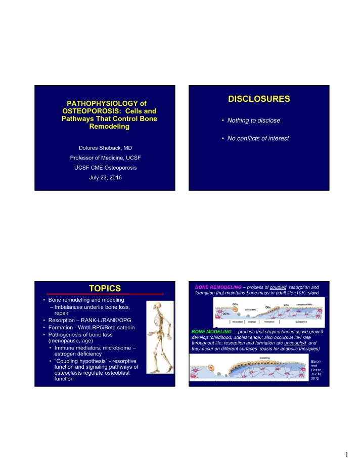
TOPICS
- Bone remodeling and modeling
– Imbalances underlie bone loss, repair
- Resorption – RANK-L/RANK/OPG
- Formation - Wnt/LRP5/Beta catenin
- Pathogenesis of bone loss
(menopause, age)
- Immune mediators, microbiome –
estrogen deficiency
- “Coupling hypothesis” - resorptive
function and signaling pathways of
- steoclasts regulate osteoblast
function
Baron and Hesse, JCEM, 2012