
SLIDE 1
3/7/2018 1
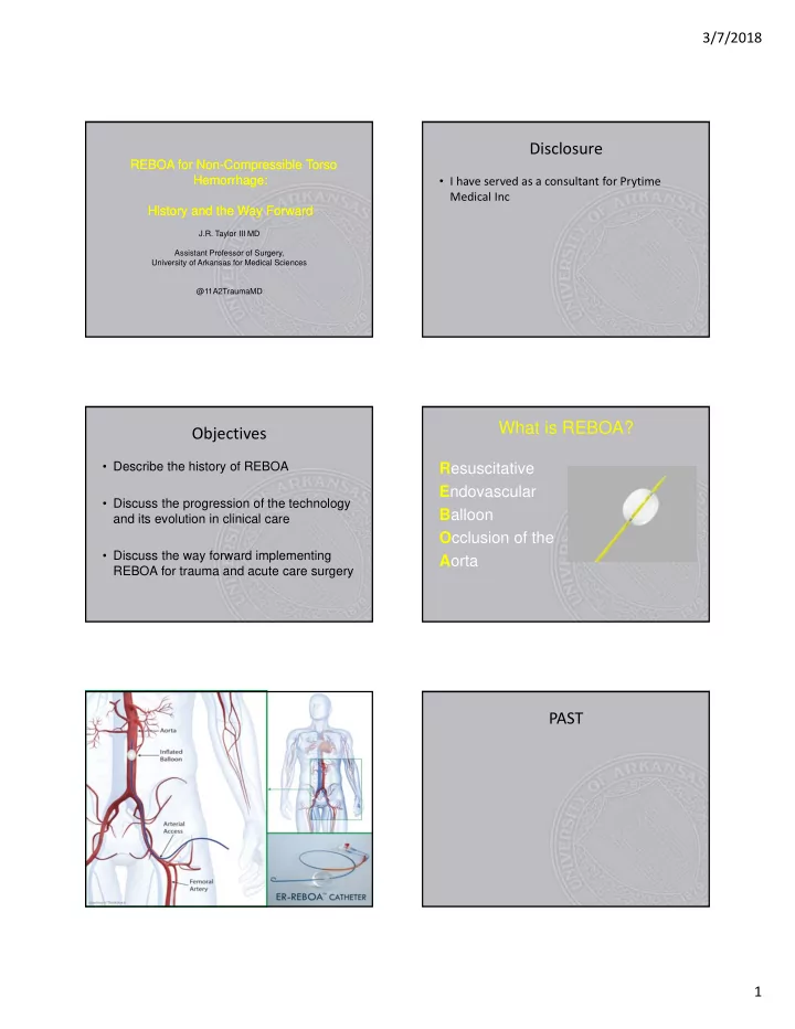
REBOA for Non-Compressible Torso Hemorrhage: History and the Way Forward REBOA for Non-Compressible Torso Hemorrhage: History and the Way Forward
J.R. Taylor III MD Assistant Professor of Surgery, University of Arkansas for Medical Sciences @11A2TraumaMD
Disclosure
- I have served as a consultant for Prytime
Medical Inc
Objectives
- Describe the history of REBOA
- Discuss the progression of the technology
and its evolution in clinical care
- Discuss the way forward implementing