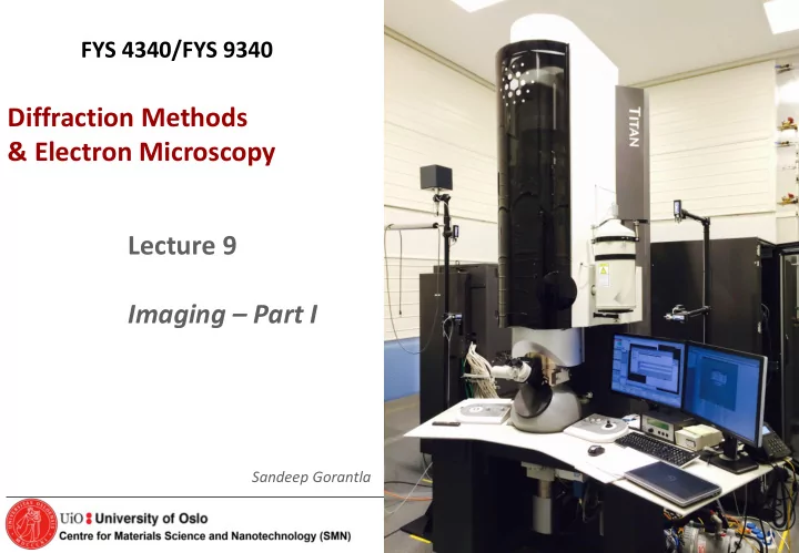

FYS 4340/FYS 9340 Diffraction Methods & Electron Microscopy Lecture 9 Imaging – Part I Sandeep Gorantla FYS 4340/9340 course – Autumn 2016 1
Imaging 2
Abbe’s principle of imaging Unlike with visible light, due to the small l, electrons can be coherently scattered by crystalline samples so the diffraction pattern at the back focal plane of the object corresponds to the sample Rays with same q converge reciprocal lattice. (inverted) 4
Abbe’s principle of imaging A diffraction pattern is always formed at the back focal plane of the objective (even in OM). To view this diffraction pattern one has to change the excitation of the intermediate lens. A higher strength projects the specimen image on the screen, a lower strength project the DP. The optical system of the TEM: The objective lens simultaneously generates the diffraction pattern and the first intermediate image. Note that the ray paths are identical until the intermediate lens, where the field strengths are changed, depending on the desired operation mode. A higher field strength (shorter focal length) is used for imaging, whereas a weaker field strength (longer focal length) is used for diffraction. Contrast enhancement requires mastering the TEM both in diffraction and imaging modes … screen 5 5
How does an image in FOCUS look like in TEM??? WHEN YOU ARE IN FOCUS IN TEM THE CONTRAST IS MINIMUM IN IMAGE (AT THE THINNEST PART OF THE SAMPLE) OVER FOCUS FOCUS UNDER FOCUS DARK NO BRIGHT FRINGE FRINGE FRINGE α 1 > α 2 FRINGES OCCURS AT EDGE DUE TO FRESNEL DIFFRACTION Courtesy : D.B. Williams & C.B. Carter, Transmission electron microscopy 6
Basic imaging mode: Bright field In the bright field (BF) mode of the TEM, an aperture is inserted into the back focal plane of the objective lens, the same plane at which the diffraction pattern is formed. The aperture allows only the direct beam to pass. The scattered electrons are blocked. Dark regions are strongly dispersing the light. This is the most common imaging technique. 7 7
Basic imaging mode: Dark field (used usually only for crystalline materials) In dark field (DF) images, the direct beam is blocked by the aperture while one or more diffracted beams are allowed to pass trough the objective aperture. Since diffracted beams have strongly interacted with the specimen, very useful information is present in DF images, e.g., about planar defects, stacking faults, dislocations, particle/grain size. 8 8
Interaction of Electrons with the specimen in TEM Electrons have both wave and particle nature Typical specimen thickness ~ 100 nm or less Scattered beam (Bragg’s scattered e - ) Direct beam (Forward scattered e - ) Bragg’s scattered e - : Coherently scattered electrons by the atomic planes in the specimen which are oriented with respect t o the incident beam to satisfy Bragg’s diffraction condition Courtesy : D.B. Williams & C.B. Carter, Transmission electron microscopy 9
Bright Field and Dark Field TEM image formation Simplified Ray diagram of TEM imaging mode Diffraction Pattern Scattered beam Scattered beam 200 nm Direct beam Direct beam Scattered beam Direct beam or Image Diffraction Plane Scattered beam Objective aperture In the image ONLY Direct BEAM ONLY Scattered BEAM 2 0 0 n m Dark Field TEM image Bright Field TEM image FYS 4340/9340 course – Autumn 2016 10
Example of Bright Field and Dark Field TEM Bright Field TEM image Dark Field TEM image Objective aperture Low image contrast More image contrast More image contrast Cu 2 O Cu 2 O Cu 2 O ZnO ZnO ZnO 1 0 0 n m 1 0 0 n m 11
Contrast mechanisms The image contrast originates from: Amplitude contrast • Mass - The only mechanism that generates contrast for amorphous materials: Polymers and biological materials • Diffraction - Only exists with crystalline materials: metals and ceramics Phase (produces images with atomic resolution) Only useful for THIN crystalline materials (diffraction with NO change in wave amplitude): Thin metals and ceramics 12
Mass contrast • Mass contrast: Variation in mass, thickness or both • Bright Field (BF): The basic way of forming mass-contrast images • No coherent scattering Mechanism of mass-thickness contrast in a BF image. Thicker or higher-Z areas of the specimen (darker) will scatter more electrons off axis than thinner, lower mass (lighter) areas. Thus fewer electrons from the darker region fall on the equivalent area of the image plane (and subsequently the screen), which therefore appears darker in BF images. 13
Mass contrast • Heavy atoms scatter more intensely and at higher angles than light ones. • Strongly scattered electrons are prevented from forming part of the final image by the objective aperture. • Regions in the specimen rich in heavy atoms are dark in the image. • The smaller the aperture size, the higher the contrast. • Fewer electrons are scattered at high electron accelerating voltages, since they have less time to interact with atomic nuclei in the specimen: High voltage TEM result in lower contrast and also damage polymeric and biological samples 14
Mass contrast Bright field images (J.S.J. Vastenhout, Microsc Microanal 8 Suppl. 2, 2002) In the case of polymeric and biological samples, i.e., with low atomic number and similar electron densities, staining helps to increase the imaging contrast and mitigates the radiation damage. The staining agents work by selective absorption in one of the phases and tend to stain unsaturated C-C bonds. Since they contain heavy elements with a high scattering power, the stained regions appear dark in bright field. Stained with OsO 4 and RuO 4 vapors Os and Ru are heavy metals… 15 15
Mass contrast Bright field: Typical image of a stained biological material faculty.une.edu 16 16
Diffraction contrast Bright field If the sample has crystalline areas, many electrons are strongly scattered by Bragg diffraction (especially if the crystal is oriented along a zone axis with low indices), and this area appears with dark contrast in the BF image. The scattered electron beams are deflected away from the optical axis and blocked by the objective aperture, and thus the corresponding areas appear dark in the BF-image 17 17
Diffraction contrast Dark field Dark-field images occur when the objective aperture is positioned off-axis from the transmitted beam in order to allow only a diffracted beam to pass. In order to minimize the effects of lens aberrations, the diffracted beam is deflected from the optic axis, One diffracted beam is used to form the image. This is done with the same aperture which is displaced. However, as these electrons are not on the optical axis of the instrument, they will suffer from severe aberrations that will lower the resolution. If an inclined beam is used, the diffracted beam will be at the optical axis, i.e., aberration are minimized. This is not required is aberration corrected instruments. 18 18
Two-beam conditions The [011] zone-axis diffraction pattern has many planes diffracting with equal strength. In the smaller patterns the specimen is tilted so there are only two strong beams, the direct 000 on-axis beam and a different one of the hkl off- axis diffracted beams. 19
Diffraction contrast Bright field images (two beam condition) When an electron beam strikes a sample, some of the electrons pass directly through while others may undergo slight inelastic scattering from the transmitted beam. Contrast in an image is created by differences in scattering. By inserting an aperture in the back focal plane, an image can be produced with these transmitted electrons. The resulting image is known as a bright field image. Bright field images are commonly used to examine micro-structural related features. Two-beam BF image of a twinned Crystalline defects shown in a two- crystal in strong contrast. beam BF image 20
Diffraction contrast Dark field images (two beam condition) If a sample is crystalline, many of the electrons will undergo elastic scattering from the various (hkl) planes. This scattering produces many diffracted beams. If any of these diffracted beams is allowed to pass through the objective aperture a dark field image is obtained. In order to reduce spherical aberration and astigmatism and to improve overall image resolution, the diffracted beam will be deflected such that it lies parallel the optic axis of the microscope. This type of image is said to be a centered dark field image. Dark field images are particularly useful in examining micro-structural detail in a single crystalline phases. Two-beam DF image of a twinned crystal in Crystalline defects shown in a two- strong contrast. beam DF image 21
the excitation error or deviation parameter Other notation (Williams and Carter): K = k D - k I = g + s The relrod at g hkl when the beam is Dq away from the exact Bragg condition. The Ewald sphere intercepts the relrod at a negative value of s which defines the vector K = g + s . The intensity of the diffracted beam as a function of where the Ewald sphere cuts the relrod is shown on the right of the diagram. In this case the intensity has fallen to almost zero. 22
Kikuchi lines Useful to determine s… Excess Kikuchi line on G spot Deficient line in transmitted spot 23
Recommend
More recommend