differential diagnosis and objectives treatment of
play

Differential Diagnosis and Objectives Treatment of Balance - PDF document

2/10/2010 Differential Diagnosis and Objectives Treatment of Balance Disorders: Describe normal interaction of vision, The interaction of vestibular and somatosensory systems visual, vestibular and Describe differential diagnosis


  1. 2/10/2010 Differential Diagnosis and Objectives Treatment of Balance Disorders: • Describe normal interaction of vision, The interaction of vestibular and somatosensory systems visual, vestibular and • Describe differential diagnosis process for somatosensory systems vestibular dysfunction • Appraise status of vision and balance 6 th International Congress of Behavioral research Optometry April 8-11, 2010 Victoria Graham, PT, DPT, OCS, NCS Functional Definition of Balance: Content Overview Ability to: • Postural Control and Balance stand still or quietly in place (slight 12.5ºsway is normal) • Differential diagnosis of vestibular dysfunction move voluntarily – there is a limit of stability within our specific base of support • Vision loss and consequences respond automatically to external challenges and regain quiet stance – called pertubations perform these tasks under various environmental conditions Dynamic Equilibrium sensory motor Model Where am I? Role of Vestibular System in Normal Function What am I going to do? Determination of body position Choice of body movement Postural control: 1. Sensory input about head position in space Compare, select Select and adjust and combine senses muscle contractile patterns (related to gravity) and acceleration. 2. Input for appropriate motor response to Visual Vestibular Somatosensory Trunk Ankle, Eye conflicting visual/somatosensory input. system system system neck thigh head Visual control: 1. Gaze stabilization with head motion Environmental Generation of Body movement interaction 2. Head stabilization with respect to vertical
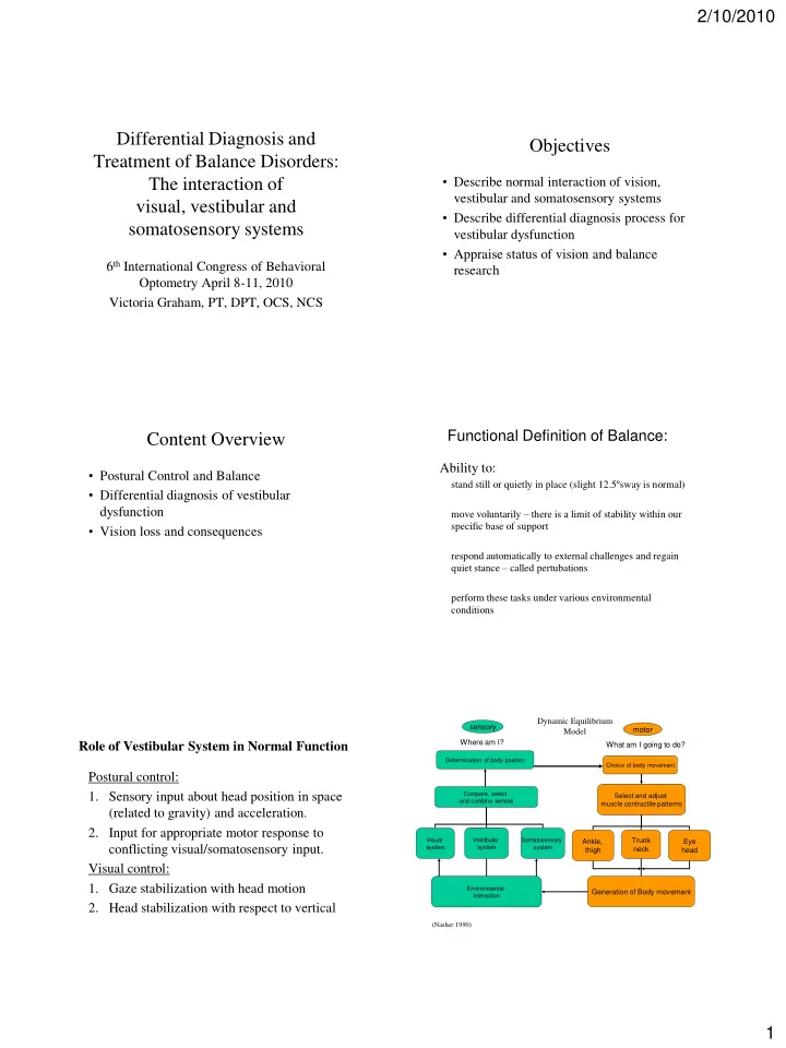
 (Nasher 1990) 1

  2. 2/10/2010 Motor strategies with perturbed stance Impact of Vision on Development of Normal Postural Control Ankle – Best strategy: Stepping • Smallest adjustment in – Largest adjustment in COM • Head control at 3 months visually driven – Slowest strategy center of mass (COM) • Efficient muscle recruitment : • Realign BOS through limb • Tonic Labyrinthine reflex supported by interaction motion – distal to proximal – Normal response to very large of eyes/head. • Energy efficient pertubations • Loss of ankle strategy • Visual mapping of hands/feet for interacting with – More likely to result in fall documented in elderly environment essential for normal function. Hip • Vision and interaction to horizon/environment – Larger adjustment in COM – Less efficient muscle recruitment: essential for developing trunk proprioception and • Slower reaction time as more muscles recruited kinesthesia. • Proximal muscle recruited first – Full body response – Normal response to larger faster pertubations Common Diagnoses involving Vestibular Causes of Dizziness System: Central: • CVA, seizure, TBI, central tumor BPPV 48% Meniere 19% Peripheral: Middle Ear 6% • BPPV, vestibular hypofunction, fistula, UVH 14% Meniere’s disease, peripheral tumor, BVH 8% Fistula 5% migraines, labyrinthitis, Differential Diagnosis: Vertigo/dizziness Vestibular Anatomy Review • Must be differentiated from non-vertiginous vertigo – Hypoxia • Labyrinth – Myocardial ischemia/cardiac arrhythmias • Vestibular afferents – Medication side effects • Efferent pathways – Infection/dehydration with electrolyte imbalance – Toxins – Hypoglycemia – Migranes – Cerebellar ataxia, basal ganglia disorders – Psychogenic 2

  3. 2/10/2010 Vestibular Labyrinth Vestibular labyrinth • Bony labyrinth • 3 semicircular canals: Contains SSC, utricle, saccule 90° from each other Filled with perilymphatic fluid (like CSF) Horizontal Anterior • Membranous labyrinth Posterior Structure suspended within body labyrinth • 2 otolith organs Supported by CT Saccule Filled with endolymph Utricle Input to Vestibular Afferents: Vestibular Afferent Nerves Hair cells that generate action potentials • SSC - stimulated by rotatory • Regular afferents fluid flow that moves cupula – Have resting firing rate of 70-100 spikes/second – Increases firing when stimulated on same side, decreases rate on Function in matched pairs opposite side PUSH-PULL mechanism – Function primarily in VOR ____________ • Irregular afferents • Otoliths – stimulated by – Typically have no resting firing rate acceleration (NOT velocity) – Function primarily in VSR motions of otoconia – Can have highly fluctuating spikes when stimulated for variable – Saccule=up/down responses – Utricle=forward/back • Both types function as matched pair in normal – both=head tilt vestibular system to give directional information. Called the push-pull mechanism Vestibular Disorder Categories Main Efferent pathways • Peripheral loss • Vestibulospinal pathway – slower velocities – Unilateral and bilateral hypofunction – Medial – to trunk/neck • Peripheral hypersensitivity – Lateral – to Lower extremities – Also has connections to limbic system – BPPV(benign paroxysmal positional vertigo) – Motion sensitivity – Migraines • Vestibulo-Occular pathway- faster velocities – Meniere’s disease (via Paramedian Pontine Reticular formation) • Central Pathology • Vestibuloccollic pathway- – Pathway disorders – Degenerative disorders Delivers vestibular information to cervical muscles 3

  4. 2/10/2010 Vestibular Disorder Categories Vestibular Disorder Categories Peripheral loss: Unilateral hypofunction: Peripheral Loss: Bilateral hypofunction – Remaining vestibular apparatus has resting – Neither vestibular apparatus is functioning firing rate ideally – Body interprets this as turning, since one side is – Patient must depend on vision and firing faster than the other (push/pull somatosensory systems mechanism) – Some evidence of neuroplastic changes are – Over time patients can adapt to the inaccurate possible, depending upon the etiology sensory information – All vestibular reflexes affected. – Many remain symptomatic for long periods – VOR remains permanently lost/damaged on one side Vestibular Disorder Categories Vestibular Disorder Categories Peripheral hypersensitivity:BPPV Peripheral hypersensitivity: Motion sensitivity (benign paroxysmal positional vertigo) • General hypersensitivity to vestibular and/or visual input. – Otoconia in utricle become dislodged • Symptoms IMMEDIATELY after mild stimulation – Move into SCC • Often people report limiting activity to avoid symptoms – Stimulate hair cells where they lie: • Can present as a vicious cycle • Free floating in SCC=canalithiasis • Responds to habituation training • Attached to cupula=cupulolithiasis • Strong link to visual system as symptoms often triggered by – Each time head is moved, the otoconia re-stimulate the SCC visual stimulus – Cause profound sensation of vertigo in certain positions (e.g.. Looking down from heights) – Etiology: head trauma, labyrinthitis, anterior vestibular artery ischemia, also spontaneous unknown etiology • Vestibular reflexes will be normal – KEY finding is LATENCY of symptoms – Vestibular Reflexes will be normal Vestibular Disorder Categories Migraine vs. Meniere’s Disease Peripheral hypersensitivity: Migraines – Common cause of episodic vertigo and Meniere’s Disease Migraine dysequilibrium – very similar symptoms to Tinnitus: high pitched Tinnitus: low-pitched, roar Meniere’s disease – difficult to differentiate May have ear fullness, Usually ear fullness or hearing • Associated with : phonophobia loss – BPPV Photophobia True spontaneous vertigo is rare; True spontaneous vertigo is – Torticollis can occur for minutes common, can occur for hours – Benign recurrent vertigo Short nap usually helps Short naps usually do not help – Motion sickness/visual stimulation of vertigo Visual auras are common Visual auras NOT common including fear of heights Motion sickness common Motion sickness NOT common – Meniere’s disease 4

Recommend


More recommend