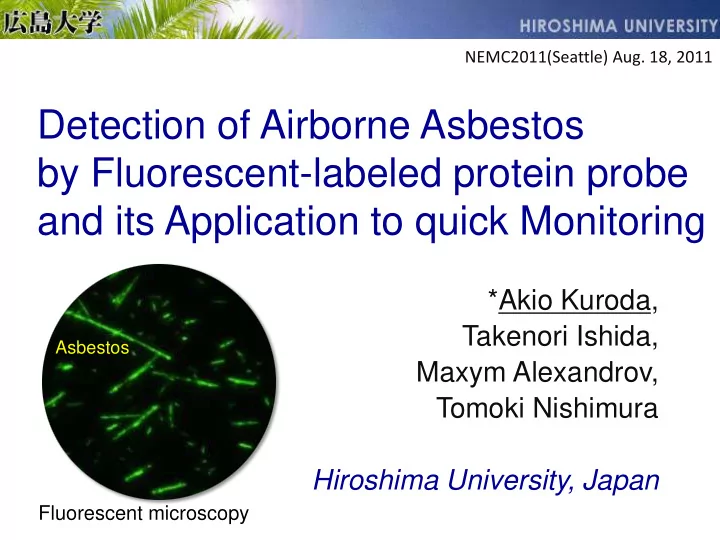
SLIDE 1 Detection of Airborne Asbestos by Fluorescent-labeled protein probe and its Application to quick Monitoring
*Akio Kuroda, Takenori Ishida, Maxym Alexandrov, Tomoki Nishimura Hiroshima University, Japan
NEMC2011(Seattle) Aug. 18, 2011
Fluorescent microscopy
Asbestos
SLIDE 2 Asbestos: silicate mineral fiber
chrysotile
(Mg6Si4O10(OH)8)
crocidolite
(Na2(Fe3+)2(Fe2+)3Si8O22(OH)2)
amosite
((Fe, Mg)7Si8O22(OH)2)
serpentine amphibole
SLIDE 3 Asbestos: widely used in construction materials
Fire retardant Slate roof Dry wall (Gypsum) Heat insulator
東日本大震災NEVER特設ページより
SLIDE 4 Asbestos: lung cancer and mesothelioma
Johnes, IARC Sci Publ. 1980;(30):637-53.1980
Asbestos exposure
1940 1950 1960 1970 1980
This has come largely as a shock to the Japanese public in 2005. Not all those deaths were workers; many of the deaths were people who lived near factories including family members of workers.
30 - 40 years latent period
mesothelioma
Japan shock 2005
SLIDE 5 Amount of asbesots in USA and Japan
750,000 500,000 250,000
37 years 32 years
USA JAPAN
Asbestos (ton)
5 million tons of asbestos are remained in Japan
SLIDE 6 Worldwide trends in the mesothelioma
Robinson and Lake, N Engl J Med 2005;353:1591-1603.
SLIDE 7
Why rapid detection method of asbestos is required?
SLIDE 8 Membrane filter asbestos
Treat with acetone vapor (transparency)
Pump Phase contrast microscopy (PCM)
Conventional method for airborne asbestos
SLIDE 9
Phase contrast microscopy (PCM)
SLIDE 10 Asbestos monitoring (Ministry of Environment)
Total fiber concentration under PCM Asbestos
under electron microscopy 【Electron microscopy wit EDX】 More than 1 fiber /L Less than 1 fiber /L OK Time consuming and expensive 【Phase contrast microscopy】
EDX
SLIDE 11 Asbestos risk at demolition site
More than 100 million tons of materials containing asbestos will be dumped until 2035. Demolition will be completed within a couple of days Rapid detection method of asbestos is required
Asbestos factory Asbestos risk at demolition site At present Past Demolition site
SLIDE 12
震災アスベスト Tagajyo city, Miyagi Pref.
Airborne asbestos from earthquake debris?
SLIDE 13
Quick detection of asbestos
asbestos Fluorescent Asbestos-binding protein
SLIDE 14
Asbestos-binding proteins
SLIDE 15 Asbestos-binding protein
Asbestos- binding protein!
actin 116 66 45 31 22 14 6 (kDa) OmpC OmpA DksA HlpA YgiW
Asbestos Mouse lung Escherichia coli Protein sources Proteins
SLIDE 16 DksA: chrysotile-binding protein
Kd = 3.5 nM 0.005 0.01 0.05 0.1 0.5 1 (mg) Asbestos (Chrysotile) Asbestos (Chrysotile)
DksA Alkaline phosphatase (AP)
Substrate purple
Enzyme asbestos detection
DksA
AsbesterTM
SLIDE 17 ペレット 1.Construction materials + AsbesterTM 3.Extraction of AsbesterTM 4.Transfer supernatant to a new tube 5.Addition
2.Centrifuge and remove supernatant
Chrysotile detection in the materials
Chrysotile content (%) A 600
Dry wall (asbestos or
non-asbestos)
Example
SLIDE 18 Materials Composition Bind Chrysotile Mg6Si4O10(OH)8
+
Antigorite Mg6Si4O10(OH)8
Mg3Si4O10(OH)2
(Mg,Fe)7Si8O22(OH)2
NaFe+3
2Fe+2 3Si8O22(OH)2
CaO-P2O5-SiO2-Al2O3
SiO2
TiO2
SiC
Mg(OH)2
Cement SiO2, CaO, etc
SiO2, CaO, etc
CaSO4
- Specificity of Asbester (DksA-AP fusion)
Indistinguishable by X-ray diffraction
SLIDE 19 Tosaka, et al., Langmuir 2010, 26(12), 9950–9955
SLIDE 20 Chrysotile
+ + + + + +
Antigorite 2-3 nm
+ +
+
How does DksA recognize asbestos?
5.5 nm
DksA
Mg-OH SiO2
Mg6Si4O10(OH)8 Mg6Si4O10(OH)8
SLIDE 21
137 Amphibole asbestos Wollastonite Aluminum silicate Titanium oxide Silicon carbide Binding
HNS: amphibole asbestos-binding protein
Amphibole asbestos Wollastonite Silicon carbide Amphibole asbestos Silicon carbide Amphibole asbestos (Amosite, Crocidolite, etc) Asbestos (amphibole asbestos)-binding region HNS 1
SLIDE 22 Specificity
Fiber DksA HNS (modified)
Asbestos
Chrysotile Bound Crocidolite Bound Amosite Bound Anthophyllite Bound Tremolite Bound Actinolite Bound
Non- asbestos
Glass wool Fine glass fiber Rockwool Fire proof fiber (RF1) Fire proof fiber (RF2) Aluminum silicate fiber Titanium potassium Slightly bound Silicon carbide whisker Bound Bound Titanium oxide whisker Wollastonite
SLIDE 23
Detection of asbestos under fluorescence microscopy
SLIDE 24
Fluorescent microscopy (FM)
SLIDE 25
Modification of protein with fluorescence
Fluorescent molecule Asbestos- binding protein Asbestos Fluorescein 491nm 521nm
SLIDE 26 Detection of airborne asbestos under FM
Filter membrane asbestos
Transparency
PCM FM
One drop of fluorescent- label protein Conven
New
SLIDE 27 Chrysotile
EDX FM SEM
(electron microscopy)
(same field)
SLIDE 28 30 nm single chrysotile fibril was detected under FM
FM
SEM
SLIDE 29
HNS-FITC(green) DksA-Cy3(red)
Double staining of asbestos
SLIDE 30
Combination of phase-contrast and fluorescence microscopy (PCM-FM fusion)
SLIDE 31
PCM
SLIDE 32
Fluorescent microscopy
Light for FM Light for PCM
SLIDE 33
①② ③ ④
SLIDE 34
①② ③ ④
SLIDE 35
①non-asbestos ②asbestos ③asbestos ④non-asbestos
SLIDE 36 Detection of Airborne Asbestos by Fluorescent-labeled protein probe and its Application to quick Monitoring
- 1. We discovered asbestos-binding proteins.
- 2. We developed a fluorescence microscopy-based
method for selective and highly sensitive detection of two different types of asbestos.
- 3. The diameter of the thinnest asbestos fibers visualized
under fluorescence microscopy was 30-35 nm.
- 4. Then we proposed PCM and FM fusion analysis.
- 5. This method could be used for on-site quick monitoring
- f airborne asbestos, for example, during demolition
work.