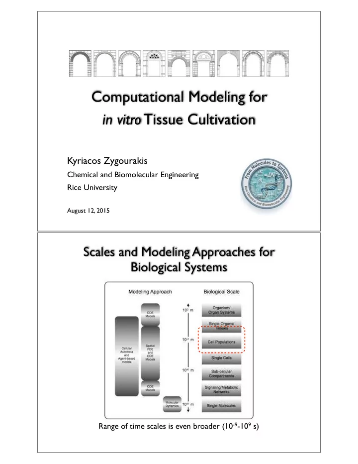

Computational Modeling for in vitro Tissue Cultivation Kyriacos Zygourakis Chemical and Biomolecular Engineering Rice University August 12, 2015 Scales and Modeling Approaches for Biological Systems Range of time scales is even broader (10 -9 -10 9 s)
Models for in vitro Tissue Cultivation Bioreactor Single Cell Design Models ? ? ? Cells migrate, proliferate and Growth differentiate Scaffold Design Factors (Biomimetics) Tissue Growth Modulated by Mass Transfer Processes • Decreased availability of nutrients (and growth factors) in the interior of 3D scaffolds at high cell densities. • The viable size of bioartificial constructs is limited to a few hundred microns due to hypoxia, nutrient insufficiency and/or waste accumulation • Large tissue replacements fail due to ( Sikavitsas et al., J. Biomed Mater. Res. , 62: 136-148, 2002 ) necrosis at the central region.
Strategies to enhance vascularization • Directing cell behavior through growth factor delivery • Using co-culturing systems • Engineering biomaterials with appropriate properties ( biomimetics ) • Incorporating microfabrication techniques • Applying mechanical stimulation ( when necessary ) (Khademhosseini et al, Tissue Eng , 2012) Initial and Boundary Conditions Are Important! Bioreactor Single Cell Design Models ? ? ? Seed cell Growth Scaffold Design distribution Factors (Biomimetics)
Competition of Dynamic Processes Cells Scaffolds Signals Cell Population Diffusion and Dynamics Uptake of Mass • Migration Dynamics Nutrients and Transfer • Proliferation Growth Factors • Differentiation ICs • Bioreactor & • Scaffold BCs • Seed cell distribution Nutrient Diffusion and Consumption Diffusion Term Nutrient consumption rate ∂ C ∂ 2 C v max C ∂ t = D e ∂ z 2 − ρ cell K m + C Diffusion Cell coefficient density Culture Media C s z = L z = 0 C s Culture Media
Nutrient Diffusion and Consumption ∂ C ∂ 2 C v max C Temporal evolution of ∂ t = D e ∂ z 2 − ρ cell K m + C nutrient concentration profiles ( ) = C s C 0 ( ) = C s 1 C L Initial 0.9 45 min 90 min 0.8 135 min Normalized Distance, z/L 180 min 0.7 L = 0.001 m 0.6 0.5 Base case parameters: 0.4 0.3 D e = 7 × 10 − 11 m 2 /s 0.2 ρ cell ⋅ v max = 8.3 × 10 − 4 mol/(m 3 ⋅ s) 0.1 C s = 5 mM 0 0 0.5 1 1.5 2 2.5 3 3.5 4 4.5 5 Nutrient concentration, mol/m 3 mM Nutrient Diffusion and Consumption ∂ C ∂ 2 C v max C Temporal evolution of ∂ t = D e ∂ z 2 − ρ cell K m + C nutrient concentration profiles ( ) = C s C 0 ( ) = C s 1 C L 0.9 0.8 Normalized Distance, z/L 0.7 L = 0.002 m Initial 0.6 60 min 0.5 120 min 180 min Base case parameters: 0.4 240 min 0.3 D e = 7 × 10 − 11 m 2 /s 0.2 ρ cell ⋅ v max = 8.3 × 10 − 4 mol/(m 3 ⋅ s) 0.1 C s = 5 mM 0 0 0.5 1 1.5 2 2.5 3 3.5 4 4.5 5 Nutrient concentration, mol/m 3 mM
Nutrient Diffusion and Consumption ∂ C ∂ 2 C v max C Temporal evolution of ∂ t = D e ∂ z 2 − ρ cell K m + C nutrient concentration profiles ( ) = C s C 0 ( ) = C s 1 C L 0.9 0.8 Normalized Distance, z/L 0.7 L = 0.004 m Initial 0.6 60 min 0.5 120 min 180 min Base case parameters: 0.4 240 min 0.3 D e = 7 × 10 − 11 m 2 /s 0.2 ρ cell ⋅ v max = 8.3 × 10 − 4 mol/(m 3 ⋅ s) 0.1 C s = 5 mM 0 0 0.5 1 1.5 2 2.5 3 3.5 4 4.5 5 Nutrient concentration, mol/m 3 mM Effect of maximum nutrient uptake rate Diffusional limitations becomes stronger when the uptake rate constant v max increases ∂ C ∂ 2 C v max C ∂ t = D e ∂ z 2 − ρ cell K m + C ( ) = C s 2 C 0 ( ) = C s 1.75 C L 1.5 1.25 Distance, mm 2 v max v max 1 Base case parameters: 0.5 v max 0.75 L = 0.002 m 0.5 D e = 7 × 10 − 11 m 2 /s ρ cell ⋅ v max = 8.3 × 10 − 4 mol/(m 3 ⋅ s) 0.25 C s = 5 mM 0 1 2 3 4 5 Nutrient concentration, mM
Effect of nutrient diffusion coefficient Diffusional limitations depend strongly on the magnitude of effective diffusivities ∂ C ∂ 2 C v max C ∂ t = D e ∂ z 2 − ρ cell K m + C ( ) = C s 2 C 0 ( ) = C s 1.75 C L 1.5 Effective diffusivity 1.25 Distance, mm 10 D e D e 1 Base case parameters: 0.1 D e 0.75 L = 0.002 m 0.5 D e = 7 × 10 − 11 m 2 /s ρ cell ⋅ v max = 8.3 × 10 − 4 mol/(m 3 ⋅ s) 0.25 C s = 5 mM 0 0 1 2 3 4 5 Nutrient concentration, mM Effect of surface nutrient concentration High surface concentrations may raise intra- tissue concentrations above the desired levels ∂ C ∂ 2 C v max C ∂ t = D e ∂ z 2 − ρ cell K m + C ( ) = C s 4 C 0 ( ) = C s 3.5 C L 3 Surface nutrient concentration 2.5 Distance, mm 10 mM 5 mM 2 Other parameters: 2.5 mM 1.5 L = 0.004 m 1 D e = 7 × 10 − 11 m 2 /s ρ cell ⋅ v max = 8.3 × 10 − 4 mol/(m 3 ⋅ s) 0.5 0 0 1 2 3 4 5 6 7 8 9 10 Nutrient concentration, mM
How to quickly estimate the extent of diffusional limitations... The Thiele Modulus Introduce dimensionless variables: u = C , ζ = z L , τ = D e t L 2 C s to obtain: ∂ τ = ∂ 2 u ∂ u u ∂ ζ 2 − φ 2 β + u 0< ζ <1, τ >0 ( ) = u 0 ζ ( ) ( ) = u 1, τ ( ) = 1 and u ζ ,0 u 0, τ ρ cell ⋅ v max Thiele modulus= Consumption rate φ = L D e ⋅ C s Diffusion rate β = K m C s
The Thiele Modulus 1 φ = 0.77 0.9 Normalized nutrient concentration, C/C s 0.8 φ = 1.54 0.7 ϕ ↑ 0.6 ⇓ 0.5 φ = 3.08 penetration 0.4 depth ↓ 0.3 φ = 6.15 0.2 0.1 φ = 12.3 0 0 0.1 0.2 0.3 0.4 0.5 0.6 0.7 0.8 0.9 1 Normalized Distance, z/L 3D Models of Ewing Sarcoma Tumors ρ cell ⋅ v max φ = L D e ⋅ C s C s ↑ ⇒ ϕ ↓ ⇒ penetration depth ↑ SEM micrograph of human EWS cells seeded in electrospun 3D PCL scaffold Fong et al, PNAS (2013) Response of human EWS cells to doxorubicin
3D Models of Ewing Sarcoma Tumors ρ cell ⋅ v max φ = L D e ⋅ C s v max ↓ ⇒ ϕ ↓ ⇒ penetration depth ↑ SEM micrograph of human EWS cells seeded in electrospun 3D PCL scaffold Fong et al, PNAS (2013) Response of human EWS cells to doxorubicin 3D Models of Ewing Sarcoma Tumors ρ cell ⋅ v max φ = L D e ⋅ C s ρ cell ↑ ⇒ ϕ ↑ ⇒ penetration depth ↓ SEM micrograph of human EWS cells seeded in electrospun 3D PCL scaffold Fong et al, PNAS (2013) Response of human EWS cells to doxorubicin
How can we overcome diffusional limitations? Perfusion Bioreactor Systems Continuous flow of media through the scaffold Bancroft, Sikavitsas and Mikos, Tissue Engineering , 9 , 549-554 (2003)
Convection, Dispersion and Consumption C 0 Assumptions: z = 0 • Axial (1-D) flow z = L • No concentration variations in radial direction • Uniform cell concentration Convection Dispersion Consumption ∂ C ∂ C ∂ z = D e , z ∂ 2 C ∂ z 2 − ρ cell V max C ∂ t + v z K m + C ∂ C ( ) at z = 0 ∂ z = v z C 0 − C Boundary conditions: D e , z ∂ C ∂ z = 0 at z = L Convection, Dispersion and Consumption Convection Dispersion Consumption ∂ u ∂ τ + Pe ∂ u ∂ ζ = ∂ 2 u u ∂ ζ 2 − φ 2 β + u Thiele modulus Peclet (Damkoeler number) Number ∂ u ( ) ∂ ζ = Pe u − 1 at ζ = 0 Boundary conditions: ∂ u ∂ ζ = 0 at ζ = 1 Dimensionless variables: u = C , ζ = z L , τ = D z t L 2 C b Dimensionless numbers: φ 2 = L 2 ρ cell ⋅ v max , Pe = L ⋅ V z , β = K m D z ⋅ C b D z C b
Will perfusion overcome the diffusional limitations? Pure Diffusion ∂ C ∂ 2 C Steady-state concentration profile v max C ∂ t = D e ∂ z 2 − ρ cell K m + C ( ) = C s 5 C 0 ( ) = C s 4.5 C L 4 Nutrient Concentration, mM 3.5 3 L = 2 mm 2.5 2 Base case parameters: 1.5 D e = 7 × 10 − 11 m 2 /s 1 ρ cell ⋅ v max = 8.3 × 10 − 4 mol/(m 3 ⋅ s) 0.5 C s = 5 mM 0 0 0.1 0.2 0.3 0.4 0.5 0.6 0.7 0.8 0.9 1 Normalized Distance, z/L Performance of perfusion bioreactors Perfusion can help maintain nutrient concentration ∂ u ∂ τ + Pe ∂ u ∂ ζ = ∂ 2 u u ∂ ζ 2 − φ 2 constant across the tissue construct β + u ∂ u ( ) ∂ ζ = Pe u − 1 at ζ = 0 6 Steady-state profile ∂ u ∂ ζ = 0 at ζ = 1 5 4.8 mM Nutrient Concentration, mM 6 min 4 L = 2 mm 3 1 min 2 min 3 min 4 min 5 min Base case parameters: 2 v z = 6.2 × 10 − 6 m/s 1 ρ cell ⋅ v max = 8.3 × 10 − 4 mol/(m 3 ⋅ s) 0 C 0 = 5 mM 0 0.1 0.2 0.3 0.4 0.5 0.6 0.7 0.8 0.9 1 Normalized Distance, z/L
Performance of perfusion bioreactors Perfusion can help maintain nutrient concentration ∂ u ∂ τ + Pe ∂ u ∂ ζ = ∂ 2 u u ∂ ζ 2 − φ 2 constant across the tissue construct β + u ∂ u ( ) ∂ ζ = Pe u − 1 at ζ = 0 6 ∂ u Steady-state profile ∂ ζ = 0 at ζ = 1 5 4.6 mM Nutrient Concentration, mM 12 min 4 L = 4 mm 3 2 min 4 min 6 min 8 min 10 min Base case parameters: 2 v z = 6.2 × 10 − 6 m/s 1 ρ cell ⋅ v max = 8.3 × 10 − 4 mol/(m 3 ⋅ s) 0 C 0 = 5 mM 0 0.1 0.2 0.3 0.4 0.5 0.6 0.7 0.8 0.9 1 Normalized Distance, z/L Performance of perfusion bioreactors Perfusion can help maintain nutrient concentration ∂ u ∂ τ + Pe ∂ u ∂ ζ = ∂ 2 u u ∂ ζ 2 − φ 2 fairly constant across the tissue construct β + u ∂ u ( ) ∂ ζ = Pe u − 1 at ζ = 0 6 ∂ u Steady-state profile ∂ ζ = 0 at ζ = 1 5 Nutrient Concentration, mM 4.3 mM 4 L = 8 mm 3 4 min 8 min 12 min 16 min 20 min Base case parameters: 2 v z = 6.2 × 10 − 6 m/s 1 ρ cell ⋅ v max = 8.3 × 10 − 4 mol/(m 3 ⋅ s) 0 C 0 = 5 mM 0 0.1 0.2 0.3 0.4 0.5 0.6 0.7 0.8 0.9 1 Normalized Distance, z/L
Recommend
More recommend