
SLIDE 1
COGS 105
Final Content Week: Brains and Clinical, Part I
wikipedia
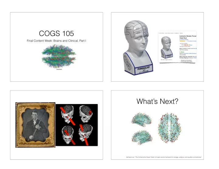
What’s Next?
Gerhard et al. “The Connectome Viewer Toolkit: An open source framework to manage, analyze, and visualize connectomes"
COGS 105 Final Content Week: Brains and Clinical, Part I wikipedia - - PowerPoint PPT Presentation
COGS 105 Final Content Week: Brains and Clinical, Part I wikipedia Whats Next? Gerhard et al. The Connectome Viewer Toolkit: An open source framework to manage, analyze, and visualize connectomes" http://www.cmtk.org/ Methods
Final Content Week: Brains and Clinical, Part I
wikipedia
Gerhard et al. “The Connectome Viewer Toolkit: An open source framework to manage, analyze, and visualize connectomes"
From reading #2
Methods
Historically, most neuroimaging studies of the human brain have employed a modular view
however, is insufficient for describing the vast set of cognitive and behavioral operations of which the brain is capable. A more appropriate approach considers which network of two or more connected and interacting regions are employed for a given function. It has not always been possible to view networks in the brain; it was not until recently that any magnetic resonance imaging (MRI) sequence was capable of discerning individual axon bundles. Traditional anatomical acquisitions used scanning protocols designed to exploit the http://www.cmtk.org/
http://journal.frontiersin.org/article/10.3389/fnhum.2013.00889/full
neuroscience for over a century has been functional localization — region X, function Y.
its underpinnings, but now backed with wicked new techniques — to study the networks of areas that underlie function.
interpretation is problematic:
and population effect modeling in relation to the underlying anatomy. NeuroImage, 61(1), 295-303.
using massive averaging and model-free analysis. Proceedings of the National Academy of Sciences, 109(14), 5487-5492.
Javier Gonzalez-Castilloa,1, Ziad S. Saadb, Daniel A. Handwerkera, Souheil J. Inatic, Noah Brenowitza, and Peter A. Bandettinia,c
aSection on Functional Imaging Methods, Laboratory of Brain and Cognition, bScientific and Statistical Computing Core, and cFunctional MRI Facility,
Institute of Mental Health, National Institutes of Health, Bethesda, MD 20892 Edited by Robert Desimone, Massachusetts Institute of Technology, Cambridge, MA, and approved February 21, 2012 (received for review December
The brain is the body’s largest energy consumer, even in the ab- sence of demanding tasks. Electrophysiologists report on-going neuronal firing during stimulation or task in regions beyond those
ical origin of consciousness remains elusive, it is argued that it emerges from complex, continuous whole-brain neuronal collabo-
continuously working and adapting to anticipate and actuate in response to the environment, over the last 20 y, task-based func- tional MRI (fMRI) have emphasized a localizationist view of brain function, with fMRI showing only a handful of activated regions in response to task/stimulation. Here, we challenge that view with evidence that under optimal noise conditions, fMRI activations extend well beyond areas of primary relationship to the task; and blood-oxygen level-dependent signal changes correlated with task-timing appear in over 95% of the brain for a simple visual stimulation plus attention control task. Moreover, we show that response shape varies substantially across regions, and that whole-brain parcellations based on those differences produce dis- tributed clusters that are anatomically and functionally meaning- ful, symmetrical across hemispheres, and reproducible across
signals beyond what is normally examined, and emphasize both the pervasiveness of false negatives, and how the sparseness of fMRI maps is not a result of localized brain function, but a conse- quence of high noise and overly strict predictive response models.
the neuronal correlates of a myriad of human behaviors. Unfortunately, if, as the previous discussion suggests, true ronal responses are continuously passing undetected in
fMRI research might be incomplete. As Lieberman and Cunningham stated previously (7), standing preoccupation with the reduction of false-posi fMRI creates a bias toward reporting only large and effects, neglecting what perhaps represents more subtle com cognitive and affective processes. Here, we explore this esis in detail and evaluate whether the sparseness of task fMRI activation maps is real or a consequence of noise lev modeling decisions. We approach this question using low fMRI time-series generated by combining unconventionall amounts of data (100 runs per subject). With these data, evaluate how regional differences in BOLD response may how distant regions collaborate during a particular task.
What Is the True Extent of BOLD Activations? Previous research
shown that if fMRI noise is reduced by time-series averaging, activation area significantly increases with number of averaged runs (8, 9). Fast increases in activation area during initial aging stages were followed by a progressive decrease in
not strong enough to attain significance with fewer trials no significant differences in hemodynamic delay from voxels were significantly active with fewer trials (8). This finding that increases in activation area could not be accounted
http://blogs.discovermagazine.com/crux/2012/04/25/does-brain-scanning-show-just-the-tip-of-the-iceberg/
Neuroskeptic http://www.nature.com/news/brain-imaging-fmri-2-0-1.10365
diffusion
“information flow.”
The connectome may be defined as the complete, point-to-point spatial connectivity of neural pathways in the brain.4 This detailed, multiscaled, and multivariate matrix is defined computationally and statistically using sophisticated in vivo neuroimaging data, electrical recordings, and postmortem tissue samples to provide a detailed framework to understand the anatomically based interactions of functional regions of the brain. The connectome gives rise to population-level atlases of distributed connectivity and makes it possible to assess disruptions of connectivity in clinical samples. Demographic, genomic, and cognitive/ behavioral data can be superimposed on the connectome to permit inferences concerning genetic and other influences on connectedness.5, 6 Information concerning connectivity is
reading #2
Bota et al., “Architecture of the cerebral cortical association connectome underlying cognition”
Cortical subplate Very strong Strong Moderate/strong Moderate Weak/moderate Weak Very Weak Not present Unknown (no data) Cortical plate Sensory-motor cortex Polymodal association cortex Exists
FROM TO
MOp MOs SSp SSs VISC ILA GU MOB AOB AON TTd TTv PIR TR PAA NLOT COAa COApl COApm AUDp AUDd AUDv VISlla VISal VISam VISli VISll VISlm VISpl VISp VISrl ACAd ACAv PL ORBl ORBm ORBv ORBvl AId AIv AIp RSPd RSPagl RSPv RSPv-a RSPv-b/c PTLp TEa ECT PERI ENTl ENTm ENTmv PRE POST PAR SUBd SUBv CA1d CA1v CA2 CA3 DG IG CLA EPd EPv LA BLAa BLAp BMAa BMAp PA MOp MOs SSp SSs VISC ILA GU MOB AOB AON TTd TTv PIR TR PAA NLOT COAa COApl COApm AUDp AUDd AUDv VISlla VISal VISam VISli VISll VISlm VISpl VISp VISrl ACAd ACAv PL ORBl ORBm ORBv ORBvl AId AIv AIp RSPd RSPagl RSPv RSPv-a RSPv-b/c PTLp TEa ECT PERI ENTl ENTm ENTmv PRE POST PAR SUBd SUBv CA1d CA1v CA2 CA3 DG IG CLA EPd EPv LA BLAa BLAp BMAa BMAp PASometimes referred to as “graph theory.”
modeling of cognition.
the structure of networks.
called an “edge.” We can analyze the structure of the network and its functional implications.
From Watts & Strogatz
clustering (C): “two friends of yours are also friends of each other” path length (λ): getting from person A to person B in how many steps?
λ λ λ λ λ intelligence quotient (IQ) such that higher IQ is associated with a more efficient cortical network, i.e., those having a “shorter” λ.31, 43 These methods have also been applied to large populations of subjects to demonstrate that the overall efficiency of the brain decreases with age44 and changes during development.45 Additionally, graph theory constructs have been used to reveal changes in the neuroanatomical networks of subjects blind from an early age46 and changes in the functional networks of attention-deficit hyperactivity disorder (ADHD) subjects.47 Indeed, the mapping of the properties, organization, and structure of brain networks will require that new network theoretical metrics be developed as we extract still finer-grained elements worthy of characterization for healthy as well as diseased or damaged networks. Meunier et al., “Modular and hierarchically modular organization of brain networks”
http://www.openconnectomeproject.org/ http://www.humanconnectomeproject.org/data/ http://www.cmtk.org/
Results, Conclusion — that’s fine.
weaving from thesis to argument to conclusion (with background review).
Analyses, Conclusion.