challenges in colorectal cancer
play

Challenges in Colorectal Cancer Wendy L Frankel, MD Chair of - PowerPoint PPT Presentation

5/28/2016 Outline- Colorectal Cancer Tumor T staging and serosal involvement Neoadjuvant treatment and staging Lymph node N staging and tumor deposits Molecular and ancillary studies Challenges in Colorectal Cancer Wendy L


  1. 5/28/2016 Outline- Colorectal Cancer � Tumor T staging and serosal involvement � Neoadjuvant treatment and staging � Lymph node N staging and tumor deposits � Molecular and ancillary studies Challenges in Colorectal Cancer Wendy L Frankel, MD Chair of Pathology Director of GI/Liver Pathology Fellowship TNM Staging 
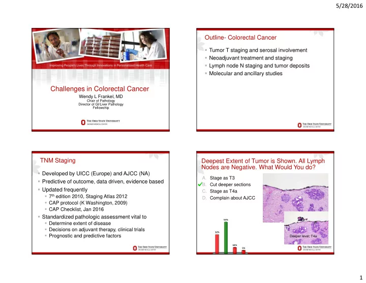
Deepest Extent of Tumor is Shown. All Lymph Nodes are Negative. What Would You do? � Developed by UICC (Europe) and AJCC (NA) A. Stage as T3 � Predictive of outcome, data driven, evidence based B. Cut deeper sections � Updated frequently C. Stage as T4a � 7 th edition 2010, Staging Atlas 2012 D. Complain about AJCC � CAP protocol (K Washington, 2009) � CAP Checklist, Jan 2016 � Standardized pathologic assessment vital to 53% � Determine extent of disease � Decisions on adjuvant therapy, clinical trials 32% � Prognostic and predictive factors Deeper level; T4a 10% 5% 1

  2. 5/28/2016 T4- Serosal Involvement CAP Cancer Staging Protocol- T4 � Absence of standard guidelines for assessing � Associated with decreased survival peritoneal involvement may contribute to � May need additional treatment underdiagnosis � Adjuvant chemotherapy recommended by ASCO for � The following findings are considered to Stage III and IV not II unless high risk features (i.e. T4) represent serosal involvement by tumor � Significant variability in reporting serosal involvement � Tumor at serosal surface with inflammatory � Studies with meticulous sampling 59% reaction, mesothelial hyperplasia, and/or � Other studies <10% erosion/ulceration � Underdiagnosed likely due to inadequate sampling � Free tumor cells on serosal surface with and not recognizing serosal penetration (up to 20%) underlying ulceration of visceral peritoneum � Both associated with decreased survival Gunderson LL, J Clin Oncol, 2010; Ludeman L, Histopathol, 2005; Shepherd NA, Gastroenterol, 1997 T4a- Serosal Surface Deeper Sections T3- close Deeper Sections, T4a 2

  3. 5/28/2016 Serosal Clefts- T4a Cytokeratin 7 is not helpful in most cases Tumor less than 1 mm; Serosal Reaction Cleft- Mesothelial Hyperplasia T3 vs. T4a � Serosal penetration under-recognized � Serosal scrape cytology 128 cases colon cancer � Peritoneal cytology + in 19% T3 (46% in T3 within 1 mm of serosal reaction) and 55% T4 Cancer � Tumor < 1 mm with reaction; T4a? � Fibroinflammatory, granulation tissue � Peritumoral abscess that communicates to surface Mesothelium � Hemorrhage, fibrin � Reactive mesothelial cells Panarelli, Am J Surg Pathol, 2013 3

  4. 5/28/2016 Elastic Stain- Why Consider it? Serosal Surface T3 or T4a? � T3 vs. T4a can be challenging � Clinically important Stage II � Possible surrogate for serosal invasion � Elastic in lung cancer for invasion visceral pleura, AJCC 7 th ed TNM � Subserosal elastic lamina colon � Located just deep to peritoneum � May be retracted toward front of carcinoma because of fibrosis � Not present in all cases If gross perforation, T4 Elastic Stain Elastic Stain- Helpful or Not? � Studies variable results; different stains and # slides � Challenges � Not all cases contain EL, particularly right colon � EL incomplete in many; need to ‘draw a line’ � Not practical if necessary to stain several slides � Reporting results (if you find EL+ deep T3) � Upstage or add comment? � I currently do not use it Deep T3 Elastic Lamina Invasion Shinto, Dis Col Rect, 2004; Kojima, Am J Surg Pathol, 2010; Kojima, Front Oncol, 2012; Liang, Am J Surg Pathol, 2013; Grin, Hum Pathol, 2013; Kojima, Am J Surg Pathol, 2013 4

  5. 5/28/2016 Deepest Extent of Tumor and Elastic Stain are Rectal Resections Shown. What Would You do? A. Stage as T3 � Radial margin most critical factor local B. Stage as T4a recurrence in rectal C. Cut deeper sections cancer (< 1mm) D. Complain about both AJCC � No 4a in non- and Elastic stains serosalized rectum 50% 47% Deeper level; T4a 2% 1% A. B. C. D. Tumor Regression Grade Neoadjuvant Chemoradiation � For advanced rectal cancer; T3-T4 and/or LN+ Description Tumor Regression Score � Improved resectability/reduced local recurrence No viable cancer cells 0 (complete response) � Associated with significant tumor response, Single cells or small groups of cells 1 (near complete response) downstaging, better prognosis � Several grading systems, modified Ryan Residual cancer with evident tumor 2 (partial response) suggested regression, but more than single cells or small groups of cells � Evaluate in tumor � Not LN or other metastatic site Extensive residual cancer with no 3 (poor or no response) evident regression � Acellular mucin likely represent treated cancer � Do not use to classify T or N 5

  6. 5/28/2016 Tumor Regression Grade Mucin Pools Tumor cells 0 (Complete Response) 2 (Partial Response) Acellular 1 (Near Complete Response) 3 (Poor Response) Mucin Pools Deeper than Malignant Cells All Other 14 Lymph Nodes are not Involved, What is the N Stage? Still ypT2 A. N0 B. N1 C. N1a D. N1 with comment Mucin in perirectal fat Still ypT2, Radial Margin- R0 57% ypT2 16% 14% 13% Mucin at radial margin Don’t upstage T or call + radial margin A. B. C. D. 6

  7. 5/28/2016 Muddy the Waters Tumor Deposits What Counts as a Lymph Node? � Nodules without residual nodal tissue (recognized 1935) � AJCC 5 th edition (1997) � Size matters, 3mm � AJCC 6 th edition (2002) � Shape matters (contour) � Round smooth � Irregular � AJCC 7 th edition (2010) � Count all separately � LN, LVI or discontinuous tumor � N1c Tumor Deposits Classify this Metastasis: What Counts as a Lymph Node? A. Involved lymph node B. Tumor deposit C. Lymphovascular invasion D. Indirect spread of tumor 63% 29% There are identifiable LN < Poor reproducibility Still subjective, residual LN 3mm; data not confirmed 6% 3% A. B. C. D. 7

  8. 5/28/2016 Lymph Node (pN) Issues in Staging- Tumor Deposit and N1c Tumor Deposit � TD can be diagnosed when � Discrete foci tumor in � No residual LN is found pericolic/perirectal fat or in adjacent mesentery away from � Do not add TD to positive LN number leading edge of tumor and no � Do not use N1c if any positive LN evidence residual LN but within lymph drainage area of primary � Do not use N1c if N1(mic)- 0.2 to 2 mm carcinoma � N1c is not worse, by definition, than N1a or b � TD- Discontinuous spread, LV with extravascular extension, or � N1c appears to be at least as bad as N1 totally replaced LN � Does not change the T stage even if tumor is T1/T2, � Identifiable LVI/LN is not TD and TD is in pericolonic tissue Jin, Am J Surg Pathol, 2015 Interobserver Study LN vs. TD: 25 Tumor Deposit (7/7) Metastasis Reviewed by 7 Pathologists Lymph Node (7/7) Rock, Arch Path Lab Med, 2013 8

  9. 5/28/2016 Challenging- TD or LN? Classify this Metastasis: Tumor Deposit (4/7) Lymph Node (4/7) A. Involved lymph node B. Tumor deposit C. Lymphovascular invasion D. Indirect spread of tumor 70% No definite residual LN or vessel Moderate agreement Useful features: round, peripheral lymphocytes/follicles, thick capsule, 14% 11% possible subcapsular sinus 5% A. B. C. D. Classify this Metastasis; If there is Already 1 Lymph Nodes Positive LN, What N Stage? � Minimum number? The more the better � Fat clearing helps, not standard practice A. Involved lymph node, N1a B. Tumor deposit, N1a � AJCC TNM 7, at least 10-14; CAP, <12, regross C. Involved lymph node, N1c � No definite minimum rectum after neoadjuvant D. Tumor deposit, N1c � Many factors affect recovery � Pathologist/surgeon experience and diligence � Patient age, sex, obesity, immune response � Length colon, procedure, site, size 48% � Future- lymph node ratio? No definite residual LN so TD; Do not 32% use N1c since another positive LN; Do not add to LN count (so not N1b) Goldstein N, Am J Surg Pathol, 2002; Cserni G, J Surg Oncol, 2002; Le Voyer TE, J Clin Oncol 11% 9% 2003; Chang GJ, JNCI, 2007; Dillman RO, Cancer, 2009; Govindarajan, J Clin Onc, 2011; de Campos-Lobato, Ann Surg Oncol, 2013 A. B. C. D. 9

  10. 5/28/2016 Tumor Budding Tumor Budding � Currently not recommended AJCC or CAP checklist � Groups of up to 5 cells at the invasive front of tumor � Associated with aggressive behavior � Most evidence where it may affect therapy � Polypectomy (?resection), Stage II CRC (?adjuvant) � Variability in criteria, high and low grade, cytokeratin, intratumoral budding (ITB) � Many studies published and ongoing Jass, JCP, 2003; Lugli, Br J Can, 2010; Lugli Hum Pathol, 2011; Bosch, Endosc, 2013; Horcic, Hum Pathol, 2013; Rogers, Mod Path, 2014 38 AJCC TNM 8; Will it be Clearer? Molecular Tests and Biomarkers � +Not required; may be clinically important � Histologic features suggestive of microsatellite instability (MSI) and Lynch � CAP biomarker template- December 2014 � MSI status � Immunohistochemistry for MMR proteins � BRAF V600E analysis � KRAS mutational analysis � MLH1 promoter methylation, NRAS , PIK3CA , PTEN 10

Download Presentation
Download Policy: The content available on the website is offered to you 'AS IS' for your personal information and use only. It cannot be commercialized, licensed, or distributed on other websites without prior consent from the author. To download a presentation, simply click this link. If you encounter any difficulties during the download process, it's possible that the publisher has removed the file from their server.

Recommend


More recommend