cardiovascular system pulmonary circuit right ventricle
play

Cardiovascular System: Pulmonary circuit: right ventricle lungs - PDF document

Cardiovascular System: Pulmonary circuit: right ventricle lungs left atrium Systemic circuit: left ventricle body right atrium Arteries = away from heart Veins = toward heart Capillaries = exchange vessels in between Amy Warenda


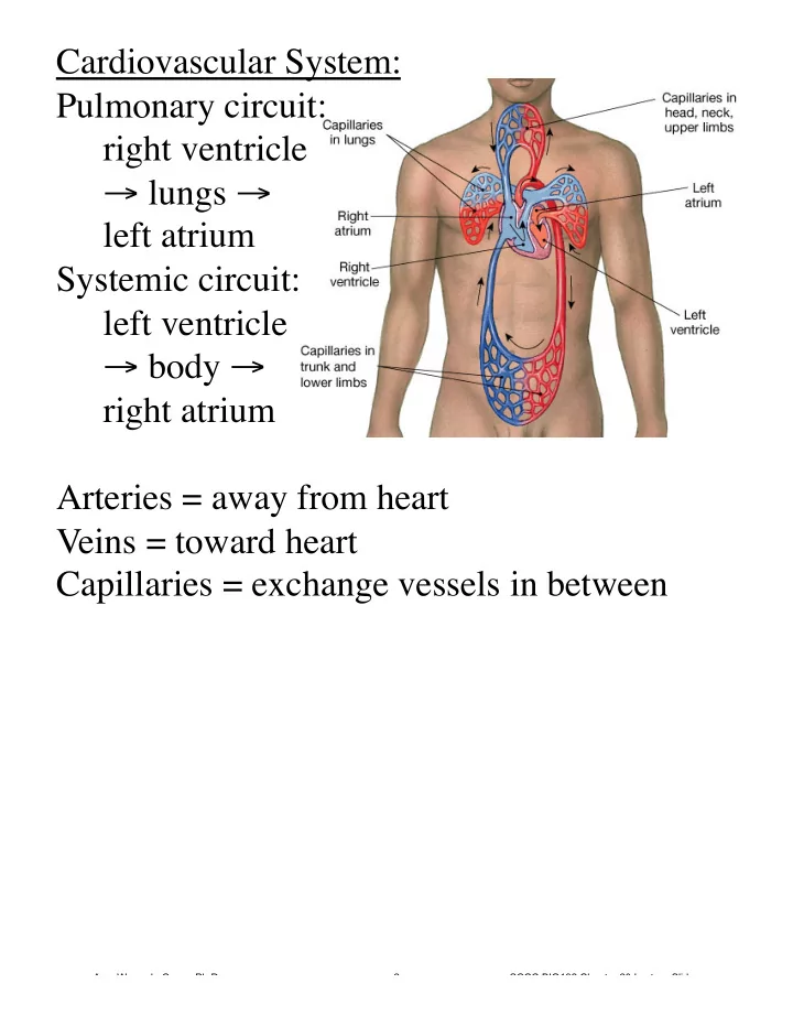
  1. Cardiovascular System: Pulmonary circuit: right ventricle → lungs → left atrium Systemic circuit: left ventricle → body → right atrium Arteries = away from heart Veins = toward heart Capillaries = exchange vessels in between Amy Warenda Czura, Ph.D. 2 SCCC BIO132 Chapter 20 Lecture Slides

  2. Heart -left of midline, between 2nd rib and 5th intercostal space, posterior to sternum, in pericardial cavity in mediastinum -heart is fist sized, < 1 lb, beats 100,000 times/day moving 8000 L blood/day -surrounded by pericardium (serous and fibrous layers) -serous membranes (visceral & parietal) secrete pericardial fluid, reduce friction Pericarditis = inflammation of pericardium, usually due to infection, causes friction Cardiac tamponade = buildup of fluid in pericardial space, restricts heart movement Amy Warenda Czura, Ph.D. 3 SCCC BIO132 Chapter 20 Lecture Slides

  3. Four Chambers: 2 Atria: -superior, thin walls, smooth posterior walls internally, pectinate muscles (ridges) anteriorly -each has expandable flap called an auricle lateral and superior -left and right separated by interatrial septum Amy Warenda Czura, Ph.D. 4 SCCC BIO132 Chapter 20 Lecture Slides

  4. 2 Ventricles: -inferior, thick walls, lined with trabeculae carneae (muscular ridges) -left and right separated by interventricular septum Amy Warenda Czura, Ph.D. 5 SCCC BIO132 Chapter 20 Lecture Slides

  5. Left ventricle 3X thicker, 5X more friction while pumping, same volume as right Left is round, right is crescent External divisions: Coronary sulcus marks division between atria and ventricles Anterior interventricular sulcus and posterior interventricular sulcus mark division between ventricles Amy Warenda Czura, Ph.D. 6 SCCC BIO132 Chapter 20 Lecture Slides

  6. Heart Wall Layers 1. Epicardium (thin) - visceral pericardium: serous membrane with loose CT attached to myocardium 2. Myocardium (thick) - cardiac muscle tissue with CT, vessels and nerves 3. Endocardium (thin) - simple squamous epithelium lining with basal lamina; continuous with endothelium of blood vessels Amy Warenda Czura, Ph.D. 7 SCCC BIO132 Chapter 20 Lecture Slides

  7. Cardiac Muscle Tissue -muscle cells = cardiocytes -uses actin and myosin sliding filaments to contract -rich in mitochondria, resists fatigue but dependent on aerobic respiration -cells connected by intercalated discs = desmosomes + gap junctions -contraction is all or none -longer contractile phase than skeletal muscle -fibrous skeleton of the heart (tough CT) acts as the tendon Amy Warenda Czura, Ph.D. 8 SCCC BIO132 Chapter 20 Lecture Slides

  8. Heart Valves (one way, prevent backflow) Atrioventricular valves - between atria and ventricles (flaps = cusps) Tricuspid valve: right side, 3 cusps Bicuspid/Mitral valve: left side, 2 cusps -cusps attached to chordae tendineae from papillary muscles on ventricle wall -contraction of papillary muscles prevent cusps opening backward during ventricle contraction -cusps hang loose when ventricles not contracting, allow ventricles to fill with blood Amy Warenda Czura, Ph.D. 9 SCCC BIO132 Chapter 20 Lecture Slides

  9. Semilunar valves - between ventricles and arteries -3 cusps -no chordae tendineae or muscles -forced open by blood from ventricular contraction -snap closed to prevent backflow Valvular heart disease - valve function deteriorates to extent that heart cannot maintain adequate circulation e.g. Rheumatic fever - childhood reaction to streptococcal infection, chronic carditis, VHD in adult Heart murmur - leaky valve (audible to Dr.) Mitral valve prolapse - murmur of left AV valve, cusps don’t close properly, blood regurgitates back into left atrium Amy Warenda Czura, Ph.D. 10 SCCC BIO132 Chapter 20 Lecture Slides

  10. Congestive heart failure - decreased pumping efficiency (diseased valves, damaged muscle), blood backs up, fluid leaks from vessels and collects in lungs and tissues Blood Flow Through The Heart (on handout) Amy Warenda Czura, Ph.D. 11 SCCC BIO132 Chapter 20 Lecture Slides

  11. Fetal Heart (adapted to bypass lungs) -Foramen ovale in right atrium, ~25% of blood bypasses directly the left atrium, closes at birth leaving scar called fossa ovalis. - Ductus arteriosus connects pulmonary trunk to aorta, ~90% of blood bypasses lungs, closes at birth leaving the ligamentum arteriosum Failure of either to close = poor oxygenation of blood, cyanosis, “blue baby syndrome” Amy Warenda Czura, Ph.D. 12 SCCC BIO132 Chapter 20 Lecture Slides

  12. Coronary circulation -heart: <1% body mass, requires 5% of blood -too thick for diffusion -coronary arteries originate at base of ascending aorta, branch to capillary beds for diffusion -blood returns via cardiac veins that empty into right atrium Amy Warenda Czura, Ph.D. 13 SCCC BIO132 Chapter 20 Lecture Slides

  13. Coronary artery disease -partial or complete block of coronary circulation, results in coronary ischemia Can lead to myocardial infarction (heart attack): heart tissue denied oxygen dies. Common symptom of CAD: angina pectoralis pain in the chest, especially during activity, as a result of the ischemia Coronary bypass surgery - use healthy veins (from legs) to create anatomoses around blockages Most people have 4 major coronary arteries: “quadruple bypass” Amy Warenda Czura, Ph.D. 14 SCCC BIO132 Chapter 20 Lecture Slides

  14. Heart Beat -1% myocardial cells autorhythmic: depolarize without neural or endocrine stimulation -depolarization transmitted to other myocardial cells through cardiac conduction system: Sinoatrial (SA) node - right atrium wall near superior vena cava Atrioventricular (AV) node - inferior portion of interatrial septum above tricuspid valve Conducting cells: AV bundle, Bundle branches, and Purkinje fibers: connect nodes and myocardium, run down interventricular septum and around apex Amy Warenda Czura, Ph.D. 15 SCCC BIO132 Chapter 20 Lecture Slides

  15. -cells of nodes cannot maintain resting membrane potential, drift to depolarization: SA node 80-100 action potentials/min (“natural pacemaker”) AV node 40-60 action potentials/min -Resting rate (sinus rhythm) ~75bpm set by SA node + parasympathetic stimulation Amy Warenda Czura, Ph.D. 16 SCCC BIO132 Chapter 20 Lecture Slides

  16. Heartbeat Chain of Events: Electrical Conduction and Contraction (on handout) Normal average heart rate= ~ 70-80bpm max = ~230bpm, but inefficient above 180 Bradycardia = heart rate slower than normal <60 Tachycardia = heart rate faster than normal >100 (Fun fact?: Lifespan theory 800 million beats) Amy Warenda Czura, Ph.D. 17 SCCC BIO132 Chapter 20 Lecture Slides

  17. Electrocardiogram (EKG) (Kardio- German) -recording of electrical events of heart (on handout) P-wave = depolarizationwave from SA node through atria ~80ms QRS complex = atrial repolarization and ventricle depolarization ~80ms T-wave = ventricle repolarization ~160ms Amy Warenda Czura, Ph.D. 18 SCCC BIO132 Chapter 20 Lecture Slides

  18. EKG used to diagnose heart problems: - P-R longer than 200ms = damage to AV node or conducting cells Total heart block = damaged AV node, no impulses transmitted through, atria and ventricles beat independently (atria fast, ventricles slow) - Large QRS = enlarged heart - Q-T longer than 380ms = coronary ischemia or myocardial damage -Cardiac arrhythmias = abnormal patterns of cardiac activity -Fibrillation = rapid, irregular, out of phase contractions due to activity in areas other than SA node: defibrillation to stop all activity so SA node can resume control Amy Warenda Czura, Ph.D. 19 SCCC BIO132 Chapter 20 Lecture Slides

  19. Normal Sinus Rhythm SA Node Not Functional, AV Node Only (Note No P Waves, Rate: 40-60 bpm) Heart Block: some P waves not conducted through AV node Ventricular fibrillation Amy Warenda Czura, Ph.D. 20 SCCC BIO132 Chapter 20 Lecture Slides

  20. Cardiac Cycle -alternating contraction and relaxation Systole = contraction, high pressure, blood gets pushed to next chamber Diastole = relaxation, low pressure, chamber fills with blood (on handout) ↑ heart rate = ↓ cycle time, ↓ diastole time = ↓ time to fill Atrial contraction only adds ~30% more to ventricles (can live with bad atria) Amy Warenda Czura, Ph.D. 21 SCCC BIO132 Chapter 20 Lecture Slides

  21. Heart Sounds “lubb” - S1: AV valves close at start of ventricular systole “dupp” - S2: semilunar valves close at start of ventricular diastole Cardiac Output CO = amount of blood pumped by each ventricle in one minute, depends on heart rate and stroke volume: CO = HR X SV SV = amount of blood pumped by ventricle Usually SV constant, change HR to increase CO as needed. HR affected by: 1. autonomic nervous input sympathetic = ↑ HR parasympathetic = ↓ HR 2. hormones 3. venous return more blood return = ↑ HR (stretch receptors activate sympathetic) 4. other factors (ions, drugs) Amy Warenda Czura, Ph.D. 22 SCCC BIO132 Chapter 20 Lecture Slides

  22. @160-180 bpm CO at max: ↑ HR = ↓ time to fill ventricles, if not full = ↓ SV and ↓ CO Conditioning can ↑ SV and ↓ HR Fit athletes can ↑ max CO by 700%, & ↓ resting HR by 50% with same CO due to ↑ SV Amy Warenda Czura, Ph.D. 23 SCCC BIO132 Chapter 20 Lecture Slides

Download Presentation
Download Policy: The content available on the website is offered to you 'AS IS' for your personal information and use only. It cannot be commercialized, licensed, or distributed on other websites without prior consent from the author. To download a presentation, simply click this link. If you encounter any difficulties during the download process, it's possible that the publisher has removed the file from their server.

Recommend


More recommend