SLIDE 1
Anatomy and Kinesiology of the Pelvic Girdle
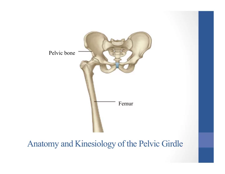
Pelvic bone Femur
SLIDE 2 Lesson Plan: 44a Anatomy and Kinesiology of the Pelvic Girdle
Breath of Arrival and Attendance
New Muscles:
- Iliopsoas
- Sartorius
- Adductors
- Deep lateral rotators
- 20 minutes:
Iliofemoral and Tibiofemoral Agonists
- 20 minutes: Reviews of muscles to be sculpted
SLIDE 3 Classroom Rules
Punctuality- everybody's time is precious:
- Be ready to learn by the start of class, we'll have you out of here on time
- Tardiness: arriving late, late return after breaks, leaving early
The following are not allowed:
- Bare feet
- Side talking
- Lying down
- Inappropriate clothing
- Food or drink except water
- Phones in classrooms, clinic or bathrooms
You will receive one verbal warning, then you'll have to leave the room.
SLIDE 4 New Muscles
Sartorius (to be sculpted today) Iliopsoas Adductor group:
- Adductor magnus
- Adductor longus
- Adductor brevis
- Pectineus
- Gracilis
Deep lateral rotators:
- Piriformis
- Obturator internus
- Gemellus superior
- Gemellus inferior
- Obturator externus
- Quadratus femoris
SLIDE 5 Sartorius
Origin:
Insertion:
Actions:
- Hip flexion
- Hip AB duction
- Knee flexion
SLIDE 6 Sartorius
Origin:
Insertion:
Actions:
- Hip flexion
- Hip AB duction
- Knee flexion
Flexion Abduction Flexion
SLIDE 7 Iliopsoas
Origin:
- Lumbar vertebrae
- Iliac fossa
Insertion:
- Lesser trochanter
- f the femur
Actions:
Psoas Major Iliacus
SLIDE 8 Iliopsoas
Origin:
- Lumbar vertebrae
- Iliac fossa
Insertion:
- Lesser trochanter of the femur
Actions:
Flexion
SLIDE 9 Adductor Group
Origin:
rami of the pubis
- Ischial ramus
- Ischial tuberosity
Insertion:
- Linea aspera
- Adductor tubercle
Actions:
Pectineus Adductor longus Gracilis Adductor brevis Adductor magnus
SLIDE 10 Adductor Group
Origin:
rami of the pubis
- Ischial ramus
- Ischial tuberosity
Insertion:
- Linea aspera
- Adductor tubercle
Actions:
AD duction
SLIDE 11 Deep Lateral Rotators
Origin:
Insertion:
- Greater trochanter
- f the femur
Actions:
(Gluteus medius, cut) (Gluteus minimus) Piriformis Superior gemellus Inferior gemellus Obturator internus Quadratus femoris
SLIDE 12 Deep Lateral Rotators
Origin:
Insertion:
- Greater trochanter
- f the femur
Actions:
Lateral Rotation
SLIDE 13 Iliofemoral Joint Muscles
Iliopsoas Sartorius Rectus femoris Tensor fascia latae Gluteals:
- Gluteus maximus
- Gluteus medius
- Gluteus minimus
Hamstrings:
- Biceps femoris
- Semitendinosus
- Semimembranosus
Adductor group:
- Adductor magnus
- Adductor longus
- Adductor brevis
- Pectineus
- Gracilis
Deep lateral rotators:
- Piriformis
- Obturator internus
- Gemellus superior
- Gemellus inferior
- Obturator externus
- Quadratus femoris
SLIDE 14
Anterior View Adductor longus TFL Sartorius Quadriceps Iliotibial band Psoas Major Iliacus Piriformis Gluteus medius Gluteus minimus Pectineus Adductor brevis Gracilis Adductor magnus Rectus femoris
SLIDE 15
Posterior View Gluteus medius TFL Gluteus maximus IT Band Vastus lateralis Adductor magnus Biceps Femoris (Plantaris) (Popliteus) Gluteus minimus Piriformis Sciatic nerve Sciatic nerve Adductor magnus Vastus lateralis Biceps femoris (Gastrocnemius) Gracilis
SLIDE 16
Agonists of the Iliofemoral Joint
SLIDE 17
Agonists of the Iliofemoral Joint
Flexion
1. Iliopsoas 2. Rectus Femoris 3. Sartorius 4. TFL
Extension
1. Gluteus Maximus 2. Hamstrings 3. Gluteus Medius (dorsal fibers)
Extension Flexion
SLIDE 18
Agonists of the Iliofemoral Joint
Abduction
1. Gluteus Medius 2. TFL 3. Gluteus Maximus (upper fibers)
Adduction
1. Adductor Group 2. Gluteus Maximus (lower fibers)
Abduction Adduction
SLIDE 19
Agonists of the Iliofemoral Joint
Medial Rotation
1. Gluteus Medius (anterior fibers) 2. TFL
Lateral Rotation
1. Gluteus Maximus 2. Deep Lateral Rotators
Lateral Rotation Medial Rotation
SLIDE 20
Agonists of the Tibiofemoral Joint
Flexion
1. Hamstrings
Extension
1. Quadriceps Femoris
Flexion Extension
SLIDE 21
Agonists of the Tibiofemoral Joint
Medial Rotation
1. Semitendinosus / Semimembranosus
Lateral Rotation
1. Biceps Femoris
Lateral Rotation Medial Rotation
SLIDE 22
SLIDE 23 Sartorius
Origin:
Insertion:
Actions:
- Hip flexion
- Hip AB duction
- Knee flexion
SLIDE 24 Sartorius
Origin:
Insertion:
Actions:
- Hip flexion
- Hip AB duction
- Knee flexion
Flexion Abduction Flexion
SLIDE 25 Biceps Femoris
Origin:
- Ischial tuberosity
- Linea aspera of femur
Insertion:
Actions:
- Flex the knee
- Extend the hip
SLIDE 26 Biceps Femoris
Origin:
- Ischial tuberosity
- Linea aspera of femur
Insertion:
Actions:
- Flex the knee
- Extend the hip
Flexion Extension
SLIDE 27 Semitendinosus
Origin:
Insertion:
(AKA: pes anserinus) Actions:
- Flex the knee
- Extend the hip
SLIDE 28 Semitendinosus
Origin:
Insertion:
(AKA: pes anserinus) Actions:
- Flex the knee
- Extend the hip
Flexion Extension
SLIDE 29 Semimembranosus
Origin:
Insertion:
- Posterior medial tibial condyle
Actions:
- Flex the knee
- Extend the hip
SLIDE 30 Semimembranosus
Origin:
Insertion:
- Posterior medial tibial condyle
Actions:
- Flex the knee
- Extend the hip
Flexion Extension
SLIDE 31
Quadriceps Femoris
SLIDE 32 Rectus Femoris
Origin:
- Anterior inferior iliac spine (ASIS)
Insertion:
- Patella
- Tibial tuberosity
Action:
- Flex the hip (femur)
- Extend the knee (tibia)
SLIDE 33 Vastus Intermedius
Origin:
- Anterior and lateral shaft of the femur
Insertion:
- Patella
- Tibial tuberosity
Action:
SLIDE 34 Vastus Medialis
Origin:
- Medial lip of linea aspera
Insertion:
- Patella
- Tibial tuberosity
Action:
SLIDE 35 Vastus Lateralis
Origin:
- Lateral lip of linea aspera
- Gluteal tuberosity
Insertion:
- Patella
- Tibial tuberosity
Action:
SLIDE 36
Quadriceps Femoris
SLIDE 37
Gastrocnemius
Origin: Posterior condyles of the femur Insertion: Posterior calcaneus Actions: Ankle plantarflexion Knee flexion
SLIDE 38
Soleus
Origin: Upper posterior tibia and fibula Insertion: Posterior calcaneus Actions: Ankle plantarflexion
SLIDE 39
Tibialis Anterior and Peroneus Longus
SLIDE 40
Tibialis Anterior
Origin: Upper 2/3 of lateral tibia Interosseous membrane Insertion: Base of 1st metatarsal Medial cuneiform (plantar) Actions: Ankle dorsiflexion Foot inversion
SLIDE 41
Tibialis Anterior
Origin: Upper 2/3 of lateral tibia Interosseous membrane Insertion: Base of 1st metatarsal Medial cuneiform (plantar) Actions: Ankle dorsiflexion Foot inversion
SLIDE 42
Tibialis Anterior
Origin: Upper 2/3 of lateral tibia Interosseous membrane Insertion: Base of 1st metatarsal Medial cuneiform (plantar) Actions: Ankle dorsiflexion Foot inversion
SLIDE 43
Tibialis Anterior
Origin: Upper 2/3 of lateral tibia Interosseous membrane Insertion: Base of 1st metatarsal Medial cuneiform (plantar) Actions: Ankle dorsiflexion Foot inversion
SLIDE 44
Peroneus Longus
Origin: Proximal 2/3 of lateral fibula Insertion: Base of 1st metatarsal Medial cuneiform (plantar) Actions: Ankle plantarflexion Foot eversion
SLIDE 45
Peroneus Longus
Origin: Proximal 2/3 of lateral fibula Insertion: Base of 1st metatarsal Medial cuneiform (plantar) Actions: Ankle plantarflexion Foot eversion
SLIDE 46
Peroneus Longus
Origin: Proximal 2/3 of lateral fibula Insertion: Base of 1st metatarsal Medial cuneiform (plantar) Actions: Ankle plantarflexion Foot eversion
SLIDE 47
Peroneus Longus
Origin: Proximal 2/3 of lateral fibula Insertion: Base of 1st metatarsal Medial cuneiform (plantar) Actions: Ankle plantarflexion Foot eversion
SLIDE 48
Tibialis Anterior and Peroneus Longus
SLIDE 49
Gluteus Maximus and Gluteus Medius
SLIDE 50
Gluteus Maximus
Origin: Posterior iliac crest Posterior sacrum and coccyx Insertion: Gluteal tuberosity of femur IT tract Actions: Extend the hip Abduct the hip Adduct the hip Laterally rotate the hip
SLIDE 51
Gluteus Medius
Origin: Upper posterior ilium Insertion: Greater trochanter of femur Actions: Abduct the hip Flex the hip Extend the hip Medially rotate the hip Laterally rotate the hip
SLIDE 52
Gluteus Maximus and Gluteus Medius
SLIDE 53
TFL and Sartorius
SLIDE 54
Tensor Fascia Latae (TFL)
Origin: Iliac crest posterior to ASIS Insertion: IT tract Actions: Abduct the hip Flex the hip Medially rotate the hip
SLIDE 55
TFL and Sartorius
SLIDE 56
Anatomy and Kinesiology of the Pelvic Girdle
Pelvic bone Femur