11/2/2011 Neuroscientists have been attracted to the puzzle of the - - PDF document

11 2 2011
SMART_READER_LITE
LIVE PREVIEW

11/2/2011 Neuroscientists have been attracted to the puzzle of the - - PDF document

11/2/2011 Neuroscientists have been attracted to the puzzle of the Cerebellum ever since Cajal. The orderly structure, the size of the cerebellum and the regularity of the neural elements demands explanation. What is the cerebellum good for?


slide-1
SLIDE 1

11/2/2011 1

Neuroscientists have been attracted to the puzzle of the Cerebellum ever since Cajal. The orderly structure, the size of the cerebellum and the regularity of the neural elements demands explanation. What is the cerebellum good for?

Ramón y Cajal 1894 Brains of vertebrates color coded by brain area. Cerebellum in orange Brains of vertebrates color coded by brain area. Cerebellum in orange All vertebrates have cerebellums, except hagfishes and lampreys. ? What does the cerebellum do? The cerebellum receives input from large areas of the cerebral cortex Directs its output to the thalamus And back to more restricted areas of cortex. The cerebellum corrects errors in each movement command. Results in motor skill. Inputs come from motor, somatosensory d l

5

and parietal cortex. Output is redirected to motor cortex Also receives input from spinal cord, and sends corrections to spinal cord. Basic circuit anatomy (the circuit is the same in all parts of the cerebellum) 1) Direct path

6

+ motor output input (sensory) deep cerebellar nucleus

slide-2
SLIDE 2

11/2/2011 2

Basic circuit anatomy (the circuit is the same in all parts of the cerebellum) 1) Direct path 2) Indirect path granule Purkinje

7

+ motor output input (sensory) deep cerebellar nucleus mossy Basic circuit anatomy (the circuit is the same in all parts of the cerebellum) 1) Direct path 2) Indirect path 3) Climbing fiber granule Purkinje climbing fiber

8

+ motor output input (sensory) deep cerebellar nucleus mossy climbing fiber We recognize cerebellums from the cellular anatomy. They have: Granule cells: receive input from mossy fibers, and send their outputs to Purkinje cells via parallel fibers Purkinje cells: the “Principal Cell” of the Cerebellum Climbing fibers: bring “error signals” from the inferior olive to ONE Purkinje cell. When a nuclear stain (Nissl) is used, the density of granule cells is

  • apparent. Purkinje cells are lightly stained, dendrites unstained. The

molecular layer looks mostly clear.

slide-3
SLIDE 3

11/2/2011 3

Vestibular Ocular Reflex (VOR)

Direct pathway from vestibular system to eye muscle

+

+ Indirect pathway via cerebellum

inferior olive

Signal from retinal “slip”

+ +

Marr, D. (1969). A theory of cerebellar

  • cortex. J. Physiol. 202, 437–470.

Albus, James A. (1971). A theory of cerebellar function. Math. Biosci. 10, 25– 61.

“Influential theories of cerebellar function have posited that highly selective or sparse coding in Granule Cells allows Purkinje cells to acquire selective responses through associative synaptic plasticity” “Similarly for cerebellum-like circuits, such as the electrosensory lobe (ELL) of mormyrid fish, Granule Cells that selectively encode specific combinations of sensory and/or motor signals could allow Purkinje-like cells to generate more specific negative images.” “Nothing is yet known about how such signals are recoded in GCs or about the significance of GC input representations for the generation of negative images.” quotes from Sawtell (2010) Neuron.

slide-4
SLIDE 4

11/2/2011 4

Learned changes in the VOR in monkeys outfitted with magnifying lenses.

Ito’s hypothesis

Visual input from superior colliculus to inferior olive: information about retinal ‘slip’ Climbing fiber input to Purkinje cell – error signal. Vestibular input from mossy fiber to granule cell to the parallel fiber in flocculo- nodular lobe of cerebellum. When co-active with error, synaptic weight of parallel fiber synapse is weakened by long term depression. Inhibition decreases and this strengthens the drive from the vestibular system increasing the gain.

Cerebellum Function: DIRECT PATHWAY: the vestibular system to the eye muscles (VOR) INDIRECT PATHWAY: vestibular collateral (mossy fiber) granule cell parallel fiber Purkinje cell (inhibits VN). ERROR SIGNAL: error in the VOR, information about image slip on the retina comes from the climbing fiber which originates in the inferior olive. (strong depolarization) Climbing fiber causes dendritic spike in the Purkinje cell s causes synaptic weights from parallel fibers to depress, weakening the inhibitory input on the vestibular neurons, thereby adjusting the gain of the eye reflex.

The Basic Architecture

1. Bell, C. C., Han, V. and Sawtell, N. B. (2008). Cerebellum-like structures and their implications for cerebellar function. Annu Rev Neurosci 31, 1-24.

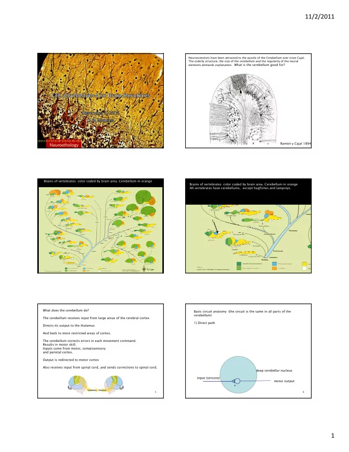
What is a Cerebellum-like structure?

Most vertebrates possess both a cerebellum and other brain structures with cerebellum-like architectures. 1) A MOLECULAR LAYER with many parallel fibers + dendrites of Purkinje-like cells. 2) A LARGE NUMBER OF GRANULE CELLS: which carry information from many central structures about the state of the

molec deep

central structures about the state of the animal: sensory information from a large number of senses, corollary discharges from motor centers (predictive of sensory input to the principle cells). 3) PRINCIPAL CELLS with SPINES. Parallel fibers terminate on spines of principal cells; or on the smooth dendrites of inhibitory stellate cells.

Cerebellum-like structures sometimes serve as adaptive sensory filters that predict sensory inputs into the deep layers using the associated parallel fiber inputs in the molecular layer.

The cerebellar-like structure is defined by the molecular layer, the principal cell, and the numerous inputs from granule cells via parallel

  • fibers. structure.
slide-5
SLIDE 5

11/2/2011 5

Cunningham's Textbook of t b D i l

25

anatomy, by Daniel John Cunningham, published in 1913 by William Wood

26

Dendritic spines

The Basic Architecture of Mormyrid ELL

1. Bell, C. C., Han, V. and Sawtell, N. B. (2008). Cerebellum-like structures and their implications for cerebellar function. Annu Rev N i 31 1 24

How does the synaptic strength change with coincident inputs?

30

slide-6
SLIDE 6

11/2/2011 6

Anti-hebbian. If the broad spike occurs after the EPSP, the synaptic weight is decreased. If the broad spike occurs before the EPSP, it strengthens. This sculpts a negative image of the expected input. causal acausal

Percentage change in excitatory postsynaptic potential (EPSP) amplitude plotted against the delay between EPSP onset and the broad spike peak during pairing. A negative delay with regard to the previous spike and a positive delay with regard to the following spike were present for each pairing. The shorter of these two delays is

  • plotted. Filled circles, significant changes; open circles, non-significant changes (at P<0.01).

animation rule

T (post-pre) broad spike before epsp broad spike after epsp amplitude +

32

delay time from epsp to broad spike

  • 500 ms + 500 ms

change in epsp a

  • ccurs when pre and

post synaptic cells

33

Natalia Caporale and Yang Dan (2008) Ann. Rev. Neurosci. fire together Gnathonemus petersii

34 35

Tim Requarth and Nathaniel B Sawtell (2011) Curr. Opin. Neurobio. Other cerebellar like structures show anti-hebbian plasticity according to slightly different learning rules.

36

slide-7
SLIDE 7

11/2/2011 7

Formation of a negative image in the ELL is just one more mechanism for removing expected inputs from sensory event.

  • Adaptation of receptors or neurons to maintained

stimuli removes responses to constant stimulus. L t l i hibiti t d l l

37

  • Lateral inhibition: remove expected mean levels
  • ver space.”

Both of these methods use simple, non-plastic cellular processes (self inhibition, intra-cellular inhibition).