
SLIDE 1
1
CEREBELLUM
Meds 5371‐ System Neuroscience
- D. L. Oliver
CEREBELLUM CEREBELLUM Anterior lobe (spinal) Posterior lobe ( - - PDF document
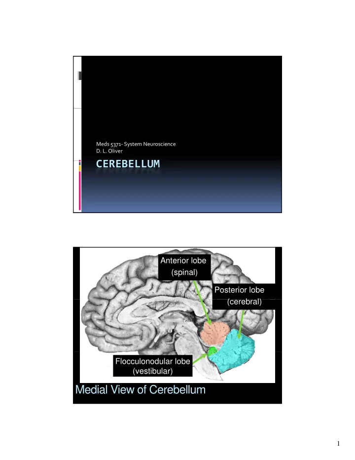
Meds 5371 System Neuroscience D. L. Oliver CEREBELLUM CEREBELLUM Anterior lobe (spinal) Posterior lobe ( (cerebral) b l) Flocculonodular lobe (vestibular) Medial View of Cerebellum 1 Ventral View of Cerebellum Flocculus Vermis
Meds 5371‐ System Neuroscience
Figure 19.1 Overall organization and subdivisions of the cerebellum (Part 4)
Superior
Middle Inferior
Inputs / Outputs with
Inputs / Outputs with
Inputs /Outputs with
Figure 19.3 Functional organization of the inputs to the cerebellum (Part 1)
Figure 19.3 Functional organization of the inputs to the cerebellum (Part 2)
INPUT – Vestibular ganglia AND vestibular nuclei
Figure 19.5 Somatotopic maps of the body surface in the cerebellum
INPUT: P ti Pontine Nucleus
Figure 19.6 Functional organization of cerebellar ouputs
Summary of Cerebellar Outputs
Figure 19.8 Functional organization of the major descending outputs from the cerebellum (Part 1)
Descending Projections
Figure 19.8 Functional organization of the major descending outputs from the cerebellum (Part 2)
Output: Fastigial nucleus projects output to vestibular nucleus Output: Flocculus, Fastigio-bulbar tract , nodulus, and vermis project directly to vestibular nucleus
Fastigial Nucleus
Fastigio-bulbar tract
Figure 19.7 Functional organization of the major ascending outputs from the cerebellum (Part 1)
Ascending Projections of Cerebellum
Figure 19.7 Functional organization of the major ascending outputs from the cerebellum (Part 2)
OUTPUT: Interposed Nuclei
Superior Cerebellar Peduncle (Brachium Conjunctivum)
OUTPUT: Dentate Nucleus
VL
Target: The VA-VL nuclei of the dorsal thalamus that project to motor cortex. Influences corticospinal tract. Other parts of dentate nucleus go to other thalamic nuclei and influence other parts of frontal lobe.
Cerebellar Cortex in Rat
A‐Purkinje B‐Basket or stellate C‐Golgi
VGAT
VGLUT1
– A‐Granule – B‐White matter
Figure 19.9 A cerebellar Purkinje neuron in a living slice from mouse cerebellum
(+) GLU
Purkinje Cell (PC)
1. Origin – Inferior olive 2. Course – Restiform body (inferior cerebellar peduncle)
Climbing fibers (CF)
(inferior cerebellar peduncle) 3. Laterality – Crossed 4. Topographical Organization - Yes 5. Destination – Purkinje cell of Cerebellar cortex
(+) GLU Granule Cell Parallel Fiber (PF) (+)
(GrC) (+) GLU Purkinje Cell (PC) Mossy Fiber (MF) Golgi Cell GABA (-) Feed-back Basket Cell GABA (-) Feed-forward
cerebellar cortex are climbing and fib mossy fibers
from cerebellar cortex inhibit the neurons of deep nuclei.
project to targets targets (outside the cerebellum) that control
major motor tracts.
Parallel Fiber (PF) Only MF & PF Active During Previously Learned Movement (+) (+) (+) (+) (+) (+) (+) (+) (+) (+) (-) (+) Mossy Fiber (MF) (+) (+) (+) (+) (+) (+) (+) (+) (+) (+)
Both CF and MF/PF Systems are Active During a New Movement
Parallel fiber fire (PF) (+) (+) (+) (+) (+) (+) (+) (+) (+) (+) (+) (+) Coincident firing
and climbing fibers causes (+) (+) Climbing fiber fires (CF) (-) (+) (+) (-) LTD at parallel fiber synapse More excitation on targets (+) from deep cerebellar nuclei Mossy Fiber (MF) (+) (+) (+) (+) (+) (+) (+) (+) (+) (+) (+) (+)