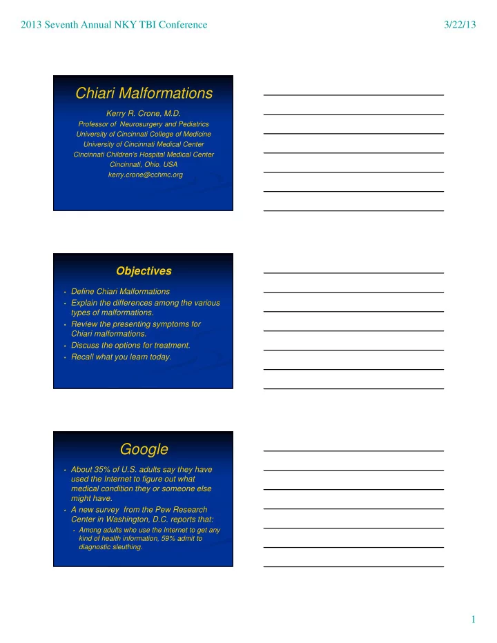
Chiari Malformations Kerry R. Crone, M.D. Professor of - - PDF document

chiari malformations
SMART_READER_LITE
LIVE PREVIEW

Chiari Malformations Kerry R. Crone, M.D. Professor of - - PDF document

2013 Seventh Annual NKY TBI Conference 3/22/13 Chiari Malformations Kerry R. Crone, M.D. Professor of Neurosurgery and Pediatrics University of Cincinnati College of Medicine University of Cincinnati Medical Center Cincinnati Childrens


slide-1
SLIDE 1

2013 Seventh Annual NKY TBI Conference 3/22/13 1

Chiari Malformations

Kerry R. Crone, M.D.

Professor of Neurosurgery and Pediatrics University of Cincinnati College of Medicine University of Cincinnati Medical Center Cincinnati Children’s Hospital Medical Center Cincinnati, Ohio. USA kerry.crone@cchmc.org

Objectives

  • Define Chiari Malformations
  • Explain the differences among the various

types of malformations.

  • Review the presenting symptoms for

Chiari malformations.

  • Discuss the options for treatment.
  • Recall what you learn today.

Google

  • About 35% of U.S. adults say they have

used the Internet to figure out what medical condition they or someone else might have.

  • A new survey from the Pew Research

Center in Washington, D.C. reports that:

  • Among adults who use the Internet to get any

kind of health information, 59% admit to diagnostic sleuthing.

slide-2
SLIDE 2

2013 Seventh Annual NKY TBI Conference 3/22/13 2

From the Internet

  • Chiari malformations are named for Hans

Chiari, an Austrian pathologist, who first identified types I-III in 1891.

  • Julius Arnold further expanded the

definition of Chiari malformation type II.

Summary Information

  • Some medical sources began using the

name Arnold-Chiari malformation.

  • Nowadays, some medical sources, use

Arnold-Chiari malformation as a broad term for all forms.

Summary Information

  • Chiari malformations have also been

known as:

  • Congenital tonsillar herniation
  • Tonsillar ectopia
  • Tonsillar descent
slide-3
SLIDE 3

2013 Seventh Annual NKY TBI Conference 3/22/13 3

The Chiari Description

  • 1891-Dr. Hans Chiari
  • Austrian pathologist
  • 17yo female with

hydrocephalus who died from typhoid fever and at autopsy her brain displayed an elongation

  • f the tonsils and medial

parts of the inferior lobes

  • f the cerebellum

The Arnold Description

  • 1894-Dr. Julius Arnold
  • German Anatomist
  • Portrayed an infant with

spina bifida and described an elongated inferior portion of the cerebellum (vermis) that covered the fourth ventricle and extended into the spinal canal.

Synthesized Terminology

  • 1907 Schwalbe and Gredig first applied

the Arnold-Chiari eponym to patients previously characterized as having the Chiari type 2 malformation.

  • Their differentiation was an improper

modification of Chiari’s original description. This eponym has continued into modern times to define rhombencephalon deformities.

slide-4
SLIDE 4

2013 Seventh Annual NKY TBI Conference 3/22/13 4

Why the Difference is Important! The Cerebellum Chiari Malformations

  • Chiari I Malformation (Most common)
  • Chiari II (Arnold-Chiari) Malformation
  • Chiari III Malformation
  • Chiari IV Malformation
  • Acquired Chiari Malformation
  • Chiari 1.5 Malformation
slide-5
SLIDE 5

2013 Seventh Annual NKY TBI Conference 3/22/13 5

Anatomic Findings in Chiari I

  • Tonsillar herniation
  • Hydrocephalus
  • Related Anomalies
  • Anatomic
  • Metabolic
  • Genetic

Anatomic Findings in Chiari II

  • Herniation of vermis,

brainstem and fourth ventricle

  • Myelomenigocele
  • Syringomyelia
  • Hydrocephalus

Chiari III Malformation

  • Posterior fossa encephalocele that

contains cerebellar and brainstem tissue with herniation through an upper cervical spina bifida.

  • Grave prognosis.
  • Must distinguish this malformation from

high cervical myelomeningoceles which may have a favorable prognosis.

slide-6
SLIDE 6

2013 Seventh Annual NKY TBI Conference 3/22/13 6

Chiari IV Malformation

  • Absent hindbrain herniation
  • Cerebellar hypoplasia or aplasia
  • Minimal function

Acquired Chiari Malformation

  • Spinal Fluid Diversion
  • Development of craniocerebral disproportion

related to chronic cranial diversion of cerebrospinal fluid. Essentially “The skull becomes to small for the brain.”

  • Herniation of cerebellar tonsils either from

chronic lumbar spinal fluid drainage related to lumboperitoneal shunts or acutely related to lumbar external drains

Chiari 1.5 Malformation

  • Transitional form of Chiari malformation

where both tonsils and brainstem are caudally descended into the cervical spine.

  • Myelomeningocele is absent.
slide-7
SLIDE 7

2013 Seventh Annual NKY TBI Conference 3/22/13 7

MR Findings in Chiari I

  • Herniation of

cerebellar tonsils

  • Absent intracranial

mass lesion

  • Tonsillar tip

configuration

  • Cine MR

Chiari I Symptoms

  • Pain
  • Occipital-cervical (lower head and neck) pain
  • Back, shoulder, and limb (arm) pain
  • Clumsiness
  • Dysphagia (difficulty swallowing)
  • Dysarthria (difficulty speaking)
  • “Dys” means bad, painful or disordered

Chiari I Clinical Findings

  • Oscillopsia
  • Esotropia
  • Crossed Eyes
  • Bradycardia
  • Slow Heart Rate
  • Apnea
  • Central
  • Hoarseness
  • Choking
  • Gagging
  • Hiccoughs
slide-8
SLIDE 8

2013 Seventh Annual NKY TBI Conference 3/22/13 8

The “Three” Chiari I Syndromes

  • Brainstem Syndrome
  • Spinal Cord Syndrome
  • Cerebellar Syndrome

Brainstem Syndrome

  • Usually from birth

through early years

  • Respiratory Irregularities
  • Nystagmus
  • Lower cranial nerve

dysfunction

  • Recurrent aspiration
  • Pneumonia
  • Reactive airway

disease

Brainstem Syndrome

slide-9
SLIDE 9

2013 Seventh Annual NKY TBI Conference 3/22/13 9

Spinal Cord Syndrome

  • Late childhood

through second decade

  • Headache
  • Scoliosis (spinal

curvature)

  • Motor/sensory losses
  • Hyporeflexia
  • Hyperreflexia

Spinal Cord Syndrome Cerebellar Syndrome

  • Usually from 18

months through adult life

  • Truncal and

appendicular ataxia

  • Sensory motor

disturbances

slide-10
SLIDE 10

2013 Seventh Annual NKY TBI Conference 3/22/13 10

Radiographic Studies

  • Magnetic Resonance Imaging
  • Pathological hindbrain herniation defined as a

distance greater than two standard deviations beyond the range of normal.

  • 6mm First Decade
  • 5mm Second and third decade
  • 4mm Fourth through eighth decade

Dysphagia

  • Dysphagia is disordered or impaired

swallowing

  • Neuromotor dysfunction
  • Developmental disabilities

Phases of Swallowing

  • Oral Phase
  • Pharyngeal Phase
  • Esophageal Phase
slide-11
SLIDE 11

2013 Seventh Annual NKY TBI Conference 3/22/13 11

Anatomy of the Medulla The Gag Reflex and Vomiting

  • The gag reflex is a normal defense

mechanism that prevents foreign bodies from entering the trachea (airway), pharynx, or larynx.

Physiology of Gagging

slide-12
SLIDE 12

2013 Seventh Annual NKY TBI Conference 3/22/13 12

Diagnosis

  • History
  • Physical Exam
  • Radiological Studies
  • MR Scan
  • Head

Spine

  • Sleep Studies (apnea)
  • Swallowing Studies (aspiration)

Treatment

  • There are only two options in the

treatment of Chiari malformations

  • Observation
  • Most common in asymptomatic patients
  • Limited published data
  • Surgery
  • Non uniform approaches

Surgical Treatment

  • Bony decompression
  • Foramen magnum (Boney opening skull

base)

  • Lamina of first cervical ring
  • Dural augmentation
  • Tonsillar coagulation
  • Fourth ventricle to subarachnoid stent
slide-13
SLIDE 13

2013 Seventh Annual NKY TBI Conference 3/22/13 13

Surgical Treatment Posterior Fossa Anatomy Bony Decompression

slide-14
SLIDE 14

2013 Seventh Annual NKY TBI Conference 3/22/13 14

Dural Band Incision Dural Band Decompression Tonsillar Pistoning

slide-15
SLIDE 15

2013 Seventh Annual NKY TBI Conference 3/22/13 15

Intraoperative Ultrasound Dural Augmentation Intraoperative Appearance

slide-16
SLIDE 16

2013 Seventh Annual NKY TBI Conference 3/22/13 16

CCHMC Chiari Database

  • Over 1200 patients referred since

1992

  • 450 patients have undergone surgical

treatment

  • Temporary Complications < 2.0%
  • 90% Patients demonstrated

Improvement

  • 5% Presenting symptoms stabilized
  • 5% No Change

Summary

  • Defined the Chiari Malformation
  • Described the differences in malformation

types.

  • Related presenting signs and symptoms.
  • Discussed options for treatment.
  • Provided an overview of surgery.

My gratitude for attention today.