
SLIDE 1
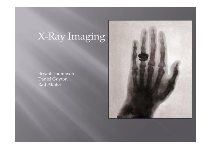
X-Ray Imaging
Bryant Thompson Bryant Thompson Daniel Guyton Rad Akhter
X-Ray Imaging Bryant Thompson Bryant Thompson Daniel Guyton Rad - - PowerPoint PPT Presentation
X-Ray Imaging Bryant Thompson Bryant Thompson Daniel Guyton Rad Akhter Easy diagnosis bone, teeth, joint etc. Fast diagnosis emergency treatments with immediate diagnosis in least invasive manner Inexpensive equipments,
Bryant Thompson Bryant Thompson Daniel Guyton Rad Akhter
Easy diagnosis bone, teeth, joint etc. Fast diagnosis emergency treatments with
Inexpensive equipments, compared to CT and
Availability majority of the facilities: hospitals,
Minimum radiation exposure radiation does not
No side effects risk of getting cancer is very small
http://benefitof.net/benefits-of-x-rays/
High voltage voltage breakdown can
Radiation exposure additional radiation
Soft-tissue imaging dense tissues appear
2D imaging limit detection ability, not
Aston, Richard. Medical Imaging Equipment Theory. 2ndnd ed. Pennsylvania: ABC Engineering Research, 2008. 38-53. Print.
Radiography 2D image, find orthopedic damage,
Mammography capture images as mamograms of
Radiography - diagnosis of Orthopedic damage http://is.sdsmt.edu/AreasofSpecialization/PreprofessionalHealthSciences/MedicalRadiographyRequirements/
pass through different parts of body creating cross-sectional images, later put together
radiation for cancer treatment
through body
angioplasty or angiography, blood flow studies, orthopedic surgery, etc.
Fluoroscopy – pacemaker leads right atrium ventricle, Pace maker implant procedure
http://www.ehow.com/list_7553903_nonmedical-uses-xrays.html
Crookes Tubes noted a fluorescent effect on barium platinocyanide screens.
first recipient of the Nobel Prize in Physics
Krohmner J.S. 1989. Radiography and Fluoroscopy, 1920 to present.
53
January 1896 Frank Austin of Dartmouth College
February 3rd, 1896 Frost brothers take image of
Frost, E. B. (1930,April). The First X-Ray Experiments in America. Dartmouth Alumni Magazine.
William D. Coolidge (1873 - 1975) In 1913 invents the Coolidge Tube, an improvement
Cathode filament made of Tungsten Became commercially available by 1917
Became commercially available by 1917
http://www.orau.org/ptp/collection/xraytubescooli dge/coolidgeinformation.htm
1896 Thomas Edison invents a modified
1912 the tilting table is made by Eugene W.
1913 Gustave Bucky creates the anti-scatter grid.
http://www.jpihealthcare.com/xray-grid
1926 Engeln Electric
1929 First rotating
ReferenceKrohmner J.S. 1989. Radiography and Fluoroscopy, 1920 to
1945 Westinghouse Electric
1948 J.W. Coltman from
1953 the Fluorex is 1953 the Fluorex is
Classic papers in modern diagnostic radiology By Adrian Thomas, Arpan K. Banerjee, Uwe Busch
Classic papers in modern diagnostic radiology By Adrian Thomas, Arpan K. Banerjee, Uwe Busch
1952 the Imperial
Used as both a
Krohmner J.S. 1989. Radiography and Fluoroscopy, 1920 to present. Radiographics. Vol. 9 no.6. pp. 1129-53
In 1983, H.Kato et al. pave way for new
The basic principle of the system is the
Eliminated the drawbacks of screen film
Digital image processing Digitization of the x-ray energy pattern by SLSL
http://www.ncbi.nlm.nih.gov/pubmed/6878707 Classic papers in modern diagnostic radiology By Adrian Thomas, Arpan K. Banerjee, Uwe Busch
2005 Scientists at UNC at Chapel Hill and Xintek,
Advantages:
Programmable electron and x-ray intensity Ultra-fine focal spot Longer lifetime Longer lifetime
http://xintek.com/newspr/news/index.htm
300 400 500 600 Price ($)
Average Cost per X-Ray Scan
100 200
"X-Ray Cost | NewChoiceHealth.com." New Choice Health. New Choice Health, Inc. Web. 30 Jan. 2012. <http://newchoicehealth.com/X-Ray-Cost>.
The cost varies greatly depending on the part of the
Small devices, such as oral X-rays can range from
Larger devices can cost anywhere from $12,000-$25,000 Larger devices can cost anywhere from $12,000-$25,000
Digital radiology machines are $50,000-$150,000, the
"Animal Insides - Digital Radiography Costs for the Veterinary Technician." Animal Insides - Welcome to Animal Insides. Animal Insides. Web. 30 Jan. 2012. <http://www.animalinsides.com/learn/the-digital-practice-integration/271- coststech.html>. "Dexis Delivers a Return on Your Investment." DEXIS: Digital X-ray for Dental Practitioners. Dexis Digital Diagnostic
In 2010, it was estimated that 182.9 million X-
That is a constant growth rate of 5.5% since
Prochaska, Gail. "IMV Reports General X-ray Procedures Growing at 5.5% per Year, as Number of Installed X-ray Units Declines." PRWeb. 11 Feb. 2011. Web. 30 Jan. 2012. <http://www.prweb.com/releases/2011/2/prweb8127064.htm>.