
SLIDE 1
1
Vascular Evaluation of the Foot
Alexander M. Reyzelman DPM Associate Professor, Dept Medicine California School of Podiatric Medicine Co-Director, UCSF Center For Limb Preservation
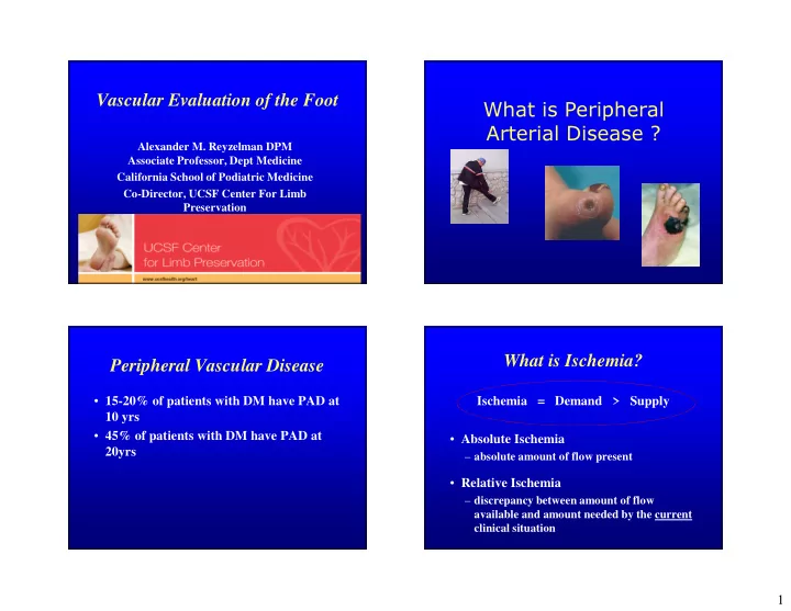
What is Peripheral Arterial Disease ?
Peripheral Vascular Disease
- 15-20% of patients with DM have PAD at
10 yrs
- 45% of patients with DM have PAD at
20yrs
What is Ischemia?
Ischemia = Demand > Supply
- Absolute Ischemia
– absolute amount of flow present
- Relative Ischemia