
SLIDE 1
1
Evaluation and Treatment of Pulmonary Arterial Hypertension
Advanced Lung Disease Course, May 2015 Teresa De Marco, MD, FACC Professor of Medicine & Surgery Director, Heart Failure and Pulmonary Hypertension Medical Director, Heart Transplantation
Presenter Disclosure Information
Evaluation and Treatment of Pulmonary Arterial Hypertension
I will not discuss off label use or investigational use in my presentation. I have financial relationships to disclose:
- Consultant for: Actelion, Gilead, United Therapeutics,
Bayer
- Research support from: United Therapeutics, Reata
- Honoraria from: Actelion, Gilead
Objectives
Review:
- Definition and classification of pulmonary
hypertension (PH) and pulmonary arterial hypertension (PAH)
- Diagnostic approach
- Treatment
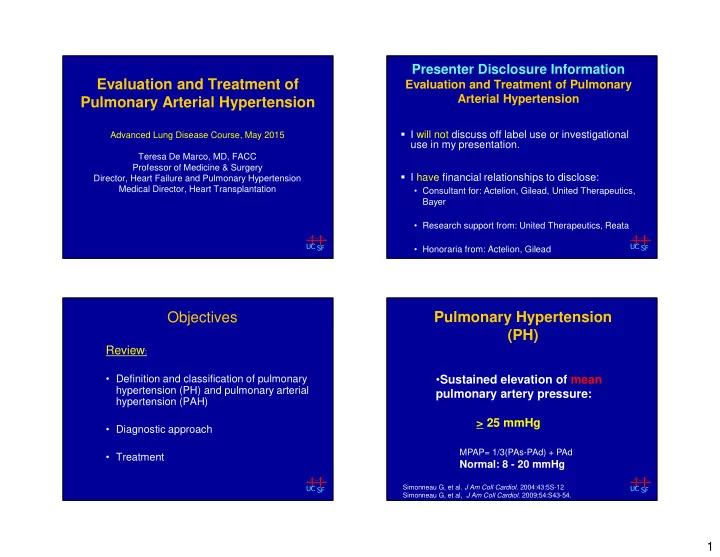
Pulmonary Hypertension (PH)
- Sustained elevation of mean
pulmonary artery pressure: > 25 mmHg
Simonneau G, et al. J Am Coll Cardiol. 2004:43:5S-12 Simonneau G, et al, J Am Coll Cardiol. 2009;54:S43-54.