Chest Radiology Highlights: Tips, Tricks and Things You Should - - PDF document

chest radiology highlights tips tricks and things you
SMART_READER_LITE
LIVE PREVIEW

Chest Radiology Highlights: Tips, Tricks and Things You Should - - PDF document

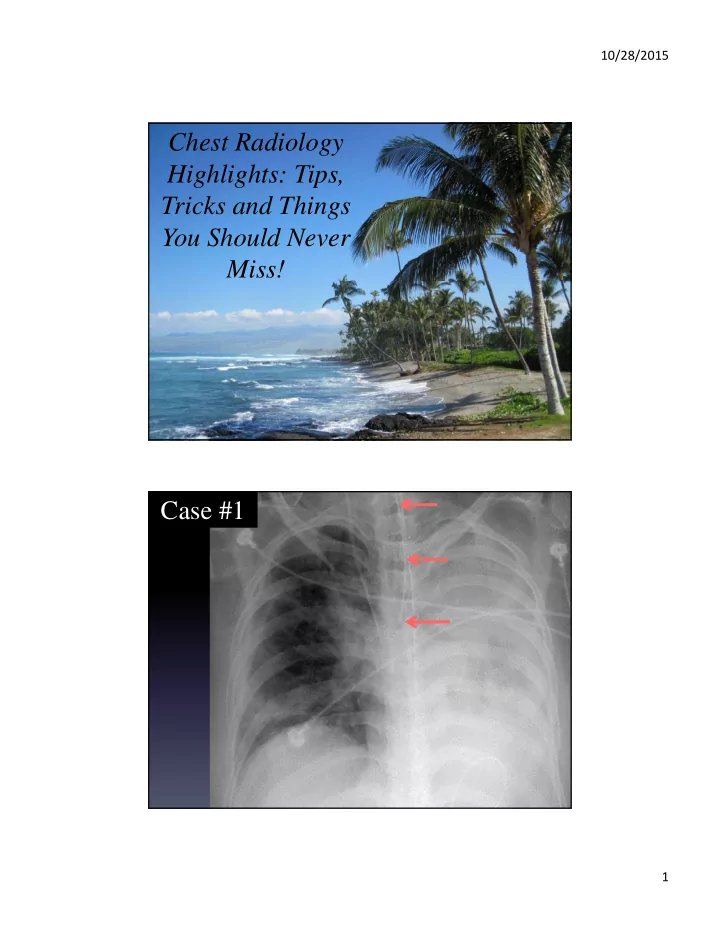
10/28/2015 Chest Radiology Highlights: Tips, Tricks and Things You Should Never Miss! Case #1 1 10/28/2015 Checklist Lines, tubes and foreign bodies Endotracheal tubes 2 10/28/2015 Airway landmarks 3 10/28/2015 3-7 cm What is


slide-1
SLIDE 1

10/28/2015 1

Chest Radiology Highlights: Tips, Tricks and Things You Should Never Miss! Case #1

slide-2
SLIDE 2

10/28/2015 2

Checklist

 Lines, tubes and foreign bodies

Endotracheal tubes

slide-3
SLIDE 3

10/28/2015 3

Airway landmarks

slide-4
SLIDE 4

10/28/2015 4 3-7 cm

What is the ideal location?

slide-5
SLIDE 5

10/28/2015 5

slide-6
SLIDE 6

10/28/2015 6

Tip position with changes in head Esophageal intubation

slide-7
SLIDE 7

10/28/2015 7

Esophageal intubation Enteric tubes

slide-8
SLIDE 8

10/28/2015 8

When is the tube post-pyloric?

GE junction 2nd duodenum 3rd duodenum 4th duodenum Ligament of Treitz 1st duodenum

slide-9
SLIDE 9

10/28/2015 9

slide-10
SLIDE 10

10/28/2015 10

slide-11
SLIDE 11

10/28/2015 11

slide-12
SLIDE 12

10/28/2015 12

slide-13
SLIDE 13

10/28/2015 13

Tension PTX s/p feeding tube removal Central lines

slide-14
SLIDE 14

10/28/2015 14 AzV SCV SCV IJ IJ RBCV LBCV SVC

Venous landmarks

The Elusive Cavoatrial Junction

slide-15
SLIDE 15

10/28/2015 15

The Elusive Cavoatrial Junction

  • 1. Junction of inferior bronchus intermedius

with lateral heart border

  • 2. Two vertebral bodies below carina
  • 3. 1-2 cm below right atrial/SVC junction

Venous landmarks

slide-16
SLIDE 16

10/28/2015 16

Variation with expiration ? location

Right posterior oblique

slide-17
SLIDE 17

10/28/2015 17

slide-18
SLIDE 18

10/28/2015 18

slide-19
SLIDE 19

10/28/2015 19 AzV SCV SCV IJ IJ RBCV LBCV SVC

slide-20
SLIDE 20

10/28/2015 20

Left SVC

slide-21
SLIDE 21

10/28/2015 21

Duplicated SVC

slide-22
SLIDE 22

10/28/2015 22

Duplicated SVC Pacemaker in left SVC

slide-23
SLIDE 23

10/28/2015 23

PICC in internal mammary vein

PICC in superior intercostal vein

slide-24
SLIDE 24

10/28/2015 24

slide-25
SLIDE 25

10/28/2015 25

Central line free in mediastinum

slide-26
SLIDE 26

10/28/2015 26

Pulmonary artery catheters

slide-27
SLIDE 27

10/28/2015 27

slide-28
SLIDE 28

10/28/2015 28

Retained surgical sponge

slide-29
SLIDE 29

10/28/2015 29

Retained catheter fragment Retained guidewire

slide-30
SLIDE 30

10/28/2015 30

Checklist

 Lines, tubes and foreign bodies

Pneumothorax

slide-31
SLIDE 31

10/28/2015 31

Checklist

 Lines, tubes and foreign bodies  Pneumothorax

Pneumothorax vs. skin fold

  • Pleural line, NOT edge
  • No vessels lateral to line
  • Line doesn’t go outside chest wall or across

midline

slide-32
SLIDE 32

10/28/2015 32 Pleural Line Air in pleura Edge Air in lung Pleural Line Air in pleura Edge Air in lung

slide-33
SLIDE 33

10/28/2015 33

Pneumothorax Where is the PTX?

slide-34
SLIDE 34

10/28/2015 34

Skin fold Skin fold

slide-35
SLIDE 35

10/28/2015 35

Overlying sheet Deep Sulcus Sign

slide-36
SLIDE 36

10/28/2015 36

Pneumothorax (subpulmonic) ? Medial PTX

slide-37
SLIDE 37

10/28/2015 37

Tension pneumothorax

slide-38
SLIDE 38

10/28/2015 38

Checklist

 Lines, tubes and foreign bodies  Pneumothorax

Case #3

slide-39
SLIDE 39

10/28/2015 39

Checklist

 Lines, tubes and foreign bodies  Pneumothorax  Mediastinal abnormalities

Normal mediastinal interfaces

Right paratracheal stripe Aorta Left pulmonary artery Aorticopulmonary window Azygoesophageal recess

slide-40
SLIDE 40

10/28/2015 40

Tuberculosis Metastatic disease (unknown primary)

slide-41
SLIDE 41

10/28/2015 41

Lung Cancer

Esophageal cancer

slide-42
SLIDE 42

10/28/2015 42

Checklist

 Lines, tubes and foreign bodies  Pneumothorax  Mediastinal abnormalities

Case #4

slide-43
SLIDE 43

10/28/2015 43

Checklist

 Lines, tubes and foreign bodies  Pneumothorax  Mediastinal abnormalities  Tough spots in the lungs

Subtle cancer #1

slide-44
SLIDE 44

10/28/2015 44

Subtle cancer #2

Subtle cancer #3

slide-45
SLIDE 45

10/28/2015 45

? pneumonia ? pneumonia

slide-46
SLIDE 46

10/28/2015 46

Normal LLL Pneumonia Normal Pleural effusion

slide-47
SLIDE 47

10/28/2015 47

Normal Nodule Normal Pott’s disease

slide-48
SLIDE 48

10/28/2015 48

Checklist

 Lines, tubes and foreign bodies  Pneumothorax  Mediastinal abnormalities  Tough spots in the lungs

Don’t forget about the bones

slide-49
SLIDE 49

10/28/2015 49

Checklist

 Lines, tubes and foreign bodies  Pneumothorax  Mediastinal lymphadenopathy  Lung nodules  Bones

Which compartment of lung is affected?

slide-50
SLIDE 50

10/28/2015 50

Alveolar Interstitial

slide-51
SLIDE 51

10/28/2015 51

Airways

Not applicable

slide-52
SLIDE 52

10/28/2015 52

Alveolar

  • Features of alveolar disease

– 1. Confluent opacities – 2. Air bronchograms – 3. Fluffy at the periphery

What produces alveolar opacities?

  • Fluid
  • Blood
  • Pus
  • Cells
slide-53
SLIDE 53

10/28/2015 53

ARDS Pulmonary edema

slide-54
SLIDE 54

10/28/2015 54

PCP Pneumonia Hemorrhage

slide-55
SLIDE 55

10/28/2015 55

Which compartment of lung is affected Interstitial

slide-56
SLIDE 56

10/28/2015 56

Interstitial Alveolar Miliary TB

slide-57
SLIDE 57

10/28/2015 57

What diseases cause small nodules?

  • Infections (fungus and tuberculosis)
  • Malignancy
  • Sarcoidosis

Miliary TB

slide-58
SLIDE 58

10/28/2015 58

Miliary fungal Sarcoidosis

slide-59
SLIDE 59

10/28/2015 59

Metastatses: lung cancer

Interstitial

slide-60
SLIDE 60

10/28/2015 60

What diseases cause linear or reticular opacities?

  • Pulmonary edema
  • Malignancy
  • Interstitial lung disease (e.g. IPF)

Lymphangitic spread of tumor

slide-61
SLIDE 61

10/28/2015 61

Edema Idiopathic pulmonary fibrosis

slide-62
SLIDE 62

10/28/2015 62

Which compartment of lung is affected?

Airways disease

  • Circular
  • Tubular
slide-63
SLIDE 63

10/28/2015 63

slide-64
SLIDE 64

10/28/2015 64

Cystic fibrosis