Using imaging to measure neural Outline: ac5vity 1. - - PDF document

using imaging to measure neural
SMART_READER_LITE
LIVE PREVIEW

Using imaging to measure neural Outline: ac5vity 1. - - PDF document

2/28/13 Using imaging to measure neural Outline: ac5vity 1. History and principles of neuroimaging 2. Review three different types of imaging Dr.


slide-1
SLIDE 1

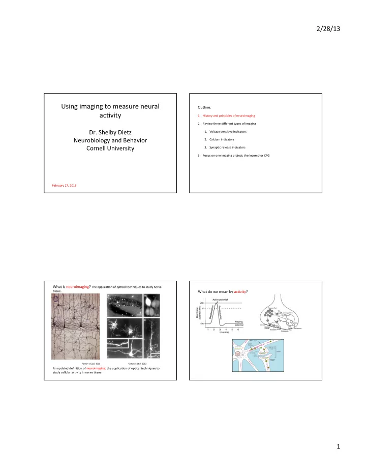
2/28/13 ¡ 1 ¡ Using ¡imaging ¡to ¡measure ¡neural ¡ ac5vity ¡ ¡

  • Dr. ¡Shelby ¡Dietz ¡

Neurobiology ¡and ¡Behavior ¡ Cornell ¡University ¡

February ¡27, ¡2013 ¡

Outline: ¡

¡

  • 1. History ¡and ¡principles ¡of ¡neuroimaging ¡
  • 2. Review ¡three ¡different ¡types ¡of ¡imaging ¡
  • 1. Voltage-­‑sensi5ve ¡indicators ¡
  • 2. Calcium ¡indicators ¡
  • 3. Synap5c ¡release ¡indicators ¡
  • 3. Focus ¡on ¡one ¡imaging ¡project: ¡the ¡locomotor ¡CPG ¡

What ¡is ¡neuroimaging? ¡The ¡applica5on ¡of ¡op5cal ¡techniques ¡to ¡study ¡nerve ¡

  • 5ssue. ¡

¡ ¡ ¡ ¡

Ramon ¡y ¡Cajal, ¡1911 ¡ ¡ ¡ ¡ ¡ ¡ ¡KeYunan ¡et ¡al. ¡2002 ¡

¡ An ¡updated ¡defini5on ¡of ¡neuroimaging: ¡the ¡applica5on ¡of ¡op5cal ¡techniques ¡to ¡ study ¡cellular ¡ac5vity ¡in ¡nerve ¡5ssue. ¡ ¡ ¡

What ¡do ¡we ¡mean ¡by ¡ac5vity? ¡

¡ ¡

slide-2
SLIDE 2

2/28/13 ¡ 2 ¡

¡

Why ¡use ¡imaging? ¡

¡ Advantages ¡of ¡imaging: ¡ ¡-­‑-­‑ ¡can ¡monitor ¡some ¡proper5es, ¡like ¡chemical ¡concentra5ons, ¡that ¡electrodes ¡ ¡ ¡can’t ¡ ¡-­‑-­‑ ¡less ¡invasive ¡than ¡an ¡electrode, ¡can ¡allow ¡for ¡long-­‑term ¡(up ¡to ¡days ¡or ¡ ¡ ¡weeks) ¡recordings ¡ ¡-­‑-­‑ ¡can ¡record ¡from ¡mul5ple ¡iden5fied ¡neurons ¡simultaneously ¡ ¡ ¡ Advantages ¡of ¡tradi5onal ¡electrophysiology ¡ ¡-­‑-­‑ ¡higher ¡fidelity, ¡can ¡resolve ¡very ¡small ¡currents ¡or ¡voltage ¡changes ¡at ¡high ¡ ¡ ¡sampling ¡rates ¡ ¡ ¡ ¡ Imaging ¡does ¡not ¡replace, ¡but ¡is ¡complementary ¡to ¡tradi5onal ¡electrophysiology ¡ ¡ ¡ ¡

First ¡op5cal ¡measurements ¡of ¡neural ¡ac5vity ¡1968 ¡

¡ ¡ ¡ ¡ ¡ ¡ ¡ ¡ ¡ ¡ ¡ ¡ ¡ ¡ Effects ¡too ¡ small ¡to ¡see ¡ individual ¡cells ¡ Func5onal ¡magne5c ¡ resonance ¡imaging ¡(fMRI): ¡ ¡ 1991 ¡ ¡ Measure ¡blood ¡oxygen ¡ ¡ ¡ ¡ ¡ ¡ ¡ ¡ ¡ ¡ ¡ ¡ ¡ ¡ ¡ ¡ To ¡record ¡the ¡ac5vity ¡of ¡individual ¡cells, ¡must ¡introduce ¡some ¡material ¡with ¡op5cal ¡ proper5es ¡that ¡change ¡more ¡drama5cally ¡in ¡response ¡to ¡changes ¡in ¡the ¡cell’s ¡

  • proper5es. ¡

First ¡op5cal ¡measurements ¡of ¡neural ¡ac5vity ¡1968 ¡

¡ ¡ ¡ ¡ ¡ ¡ ¡ ¡ ¡ ¡ ¡ ¡ ¡ ¡ ¡ ¡ ¡ ¡ ¡ ¡ ¡ ¡ ¡ ¡ ¡ ¡ ¡ ¡ ¡ What ¡can ¡be ¡used ¡to ¡ ¡ (1) detect ¡changes ¡in ¡single ¡cell ¡proper5es, ¡and ¡ ¡ (2) report ¡that ¡change? ¡ ¡ Report ¡changes ¡with ¡light ¡using ¡FLUOROPHORES ¡

First ¡op5cal ¡measurements ¡of ¡neural ¡ac5vity ¡1968 ¡

¡ ¡ ¡ ¡ ¡ ¡ ¡ ¡ ¡ ¡ ¡ ¡ ¡ ¡

slide-3
SLIDE 3

2/28/13 ¡ 3 ¡

Fluorophores ¡

¡ A ¡macromolecule ¡aYached ¡to ¡a ¡chromophore ¡that ¡can ¡fluoresce-­‑-­‑ ¡you ¡excite ¡them ¡by ¡ shining ¡light ¡of ¡one ¡wavelength ¡on ¡them, ¡and ¡they ¡convert ¡those ¡photons ¡to ¡a ¡ different ¡wavelength ¡that ¡you ¡then ¡detect. ¡

Jablonski ¡diagram ¡

If ¡you ¡aYach ¡this ¡ chromophore ¡to ¡ a ¡macromolecule ¡ that ¡changes ¡in ¡ some ¡way ¡when ¡ the ¡cell ¡changes ¡ state, ¡you ¡can ¡ signal ¡that ¡ change ¡in ¡cell ¡ state ¡with ¡a ¡ change ¡in ¡ brightness ¡or ¡ spectrum ¡ ¡ Fluorescence ¡microscope ¡configura5on ¡

What ¡are ¡some ¡of ¡the ¡characteris5cs ¡that ¡would ¡make ¡a ¡fluorophore ¡ useful ¡for ¡neural ¡imaging? ¡

¡ The ¡best ¡fluorophores ¡have ¡ ¡

  • ­‑-­‑ ¡brightness ¡

¡

  • ­‑-­‑ ¡high ¡signal-­‑to-­‑noise ¡ra5o, ¡ΔF/F ¡

¡

  • ­‑-­‑ ¡large ¡change ¡in ¡fluorescence ¡upon ¡state ¡change ¡

¡

  • ­‑-­‑ ¡fast ¡kine5cs ¡

¡

  • ­‑-­‑ ¡low ¡toxicity ¡

¡

  • ­‑-­‑ ¡long ¡dura5on ¡

¡ ¡

How ¡to ¡get ¡the ¡fluorophore ¡into ¡the ¡cells? ¡

¡ ¡ ¡ ¡ ¡ ¡ ¡ ¡ ¡ dyes: ¡almost ¡always ¡have ¡ best ¡brightness ¡and ¡kine5cs ¡ ¡ ¡ ¡ ¡ ¡ ¡ ¡ gene5cally ¡encoded: ¡ introduced ¡with ¡a ¡transgene ¡

  • r ¡virus, ¡can ¡be ¡targeted ¡to ¡

par5cular ¡cell ¡types ¡ ¡

slide-4
SLIDE 4

2/28/13 ¡ 4 ¡

The ¡big ¡problem ¡in ¡imaging: ¡signal-­‑to-­‑noise ¡

¡ Why ¡not ¡just ¡use ¡more ¡indicator, ¡drench ¡the ¡prep ¡in ¡more ¡light, ¡and ¡never ¡worry ¡about ¡ΔF/F? ¡ ¡ ¡ Problem ¡1: ¡phototoxicity– ¡light ¡kills ¡cells ¡

Hockberger ¡et ¡al. ¡1999 ¡

The ¡big ¡problem ¡in ¡imaging: ¡signal-­‑to-­‑noise ¡

¡ Why ¡not ¡just ¡use ¡more ¡indicator, ¡drench ¡the ¡prep ¡in ¡more ¡light, ¡and ¡never ¡worry ¡about ¡ΔF/F? ¡ ¡ ¡ Problem ¡2: ¡excessive ¡light ¡bleaches ¡fluorophores ¡and ¡drama5cally ¡increases ¡phototoxicity ¡

invitrogen ¡

Outline: ¡

¡

  • 1. History ¡and ¡principles ¡of ¡neuroimaging ¡
  • 2. Review ¡three ¡different ¡types ¡of ¡imaging ¡
  • 1. Voltage-­‑sensi5ve ¡indicators ¡
  • 2. Calcium ¡indicators ¡
  • 3. Synap5c ¡release ¡indicators ¡
  • 3. Focus ¡on ¡one ¡imaging ¡project: ¡the ¡locomotor ¡CPG ¡

Voltage ¡sensors ¡

¡ Light-­‑emikng ¡molecules ¡embedded ¡in ¡the ¡cell ¡membrane ¡that ¡signal ¡changes ¡in ¡ membrane ¡voltage. ¡ ¡ Dyes: ¡RH ¡414 ¡ Gene5c: ¡FlaSh, ¡SPARC ¡ ¡ ¡ ¡ ¡ ¡ ¡ ¡ ¡ ¡ ¡ ¡

From ¡Miller ¡et ¡al. ¡2012 ¡ ¡From ¡Homma ¡et ¡al. ¡2009 ¡

slide-5
SLIDE 5

2/28/13 ¡ 5 ¡

Calcium ¡sensors ¡

¡ Light-­‑emikng ¡molecules ¡floa5ng ¡in ¡the ¡cytosol ¡that ¡signal ¡changes ¡in ¡calcium ¡

  • concentra5on. ¡ ¡Increased ¡calcium ¡ ¡is ¡a ¡strong ¡indicator ¡of ¡increased ¡cellular ¡ac5vity, ¡

and ¡can ¡be ¡localized ¡to ¡a ¡single ¡synap5c ¡bouton. ¡ ¡ Dyes: ¡Fura, ¡Fluo, ¡Indo, ¡Calcium ¡Green ¡ Gene5c: ¡cameleons, ¡G-­‑CaMP, ¡Troponin-­‑C ¡ ¡ ¡

From ¡Griesbeck ¡lab ¡website, ¡Max ¡Planck ¡ From ¡Homma ¡et ¡al. ¡2009 ¡

Synap5c ¡release ¡sensors ¡

¡ Light-­‑emikng ¡molecules ¡inside ¡synap5c ¡vesicles ¡that ¡signal ¡release ¡of ¡ neurotransmiYers ¡at ¡the ¡synapse. ¡ ¡ Dyes: ¡FM-­‑143, ¡AM4-­‑64 ¡ Gene5c: ¡synaptopHluorins ¡ ¡ ¡ ¡ ¡ ¡ ¡ ¡ ¡ ¡

From ¡Burrone ¡et ¡al. ¡2007 ¡

In ¡vivo ¡imaging ¡

¡ Imaging ¡techniques ¡can ¡be ¡used ¡to ¡monitor ¡neuronal ¡ac5vity ¡in ¡living ¡animals, ¡and ¡ even ¡in ¡awake ¡and ¡behaving ¡animals. ¡ ¡

From ¡Kerr ¡and ¡Nimmerjahn ¡2012 ¡

Outline: ¡

¡

  • 1. History ¡and ¡principles ¡of ¡neuroimaging ¡
  • 2. Review ¡three ¡different ¡types ¡of ¡imaging ¡
  • 1. Voltage-­‑sensi5ve ¡indicators ¡
  • 2. Calcium ¡indicators ¡
  • 3. Synap5c ¡release ¡indicators ¡
  • 3. Focus ¡on ¡one ¡imaging ¡project: ¡the ¡locomotor ¡CPG ¡
slide-6
SLIDE 6

2/28/13 ¡ 6 ¡

Miracle ¡Mike ¡the ¡headless ¡chicken ¡survived ¡for ¡18 ¡ months ¡amer ¡a ¡botched ¡butchering ¡

¡ ¡ ¡ ¡ ¡ The ¡isolated ¡spinal ¡cord ¡CPG ¡is ¡a ¡key ¡area ¡of ¡ study ¡because ¡the ¡defined ¡inputs ¡(central ¡ drive) ¡and ¡output ¡(paYerned ¡firing ¡of ¡ventral ¡ motor ¡roots) ¡allows ¡us ¡to ¡probe ¡the ¡circuitry ¡

  • f ¡a ¡mammalian ¡neural ¡network. ¡

¡ ¡ ¡ ¡

Goulding ¡2009 ¡

Closeup ¡of ¡an ¡imaging ¡project: ¡ studying ¡individual ¡cells ¡in ¡the ¡ locomotor ¡CPG ¡

Is ¡the ¡mammalian ¡locomotor ¡CPG ¡driven ¡by ¡pacemaker ¡cell ¡class? ¡ ¡

Two ¡types ¡of ¡CPGs: ¡ ¡rhythm ¡driven ¡by ¡a ¡class ¡of ¡pacemaker ¡cells, ¡or ¡deriving ¡from ¡ emergent ¡proper5es ¡of ¡excita5on ¡and ¡inhibi5on ¡among ¡many ¡cell ¡types ¡

Marder ¡and ¡Bucher ¡2001 ¡ Hinckley ¡et ¡al. ¡2005 ¡ Hinckley ¡et ¡al. ¡2005 ¡

¡ ¡ ¡ ¡ One ¡class ¡of ¡interneurons ¡in ¡the ¡locomotor ¡CPG, ¡ Hb9, ¡fit ¡many ¡of ¡the ¡criteria ¡for ¡a ¡pacemaker ¡cell ¡in ¡ a ¡slice ¡or ¡hemicord: ¡ ¡ ¡ ¡-­‑-­‑ ¡firing ¡in ¡5me ¡with ¡hemicord ¡locomotor ¡rhythm ¡ ¡ ¡ ¡ ¡ ¡ ¡-­‑-­‑ ¡endogenous ¡oscilla5ons ¡ ¡ ¡-­‑-­‑ ¡intrinsic ¡proper5es ¡like ¡rebound ¡excita5on ¡ ¡ ¡

Proper5es ¡of ¡puta5ve ¡pacemaker ¡cell ¡class, ¡Hb9 ¡

To ¡determine ¡the ¡true ¡role ¡of ¡Hb9 ¡INs, ¡we ¡must ¡study ¡their ¡group ¡ac5vity ¡in ¡an ¡intact ¡spinal ¡cord. ¡

Wilson ¡et ¡al. ¡2005 ¡

Problem ¡1: ¡how ¡to ¡record ¡from ¡the ¡cells ¡while ¡an ¡animal ¡is ¡walking? ¡ ¡ Solu5on: ¡use ¡fic5ve ¡locomo5on ¡

¡

  • ¡in ¡vitro ¡isolated ¡neonatal ¡mouse ¡spinal ¡cord ¡can ¡generate ¡alterna5ng ¡bursts ¡in ¡the ¡

ventral ¡motor ¡roots ¡

  • ¡can ¡be ¡elicited ¡by ¡applica5on ¡of ¡excitatory ¡neurotransmiYers ¡(e.g. ¡glutamate), ¡

neuromodulators ¡(e.g. ¡5-­‑HT), ¡or ¡by ¡a ¡variety ¡of ¡electrical ¡s5muli ¡ ¡ ¡ ¡ ¡ ¡ ¡ ¡ ¡ ¡ ¡ ¡ ¡

Kiehn ¡et ¡al. ¡2010 ¡ Kiehn ¡et ¡al. ¡2010 ¡ Gordon ¡and ¡Whelan ¡2006 ¡

Drug ¡induced ¡ ¡ ¡Electrically ¡induced ¡

slide-7
SLIDE 7

2/28/13 ¡ 7 ¡

Problem ¡2: ¡the ¡cells ¡are ¡too ¡deep ¡and ¡too ¡numerous ¡to ¡record ¡with ¡electrodes ¡ ¡ Solu5on: ¡Calcium ¡imaging! ¡ ¡Load ¡Hb9 ¡cells ¡with ¡Ca ¡dye. ¡

2-­‑photon ¡objec5ve ¡

image ¡iden5fying ¡GFP+ ¡cells ¡ ¡ ¡ image ¡iden5fying ¡Fluo-­‑3 ¡labeled ¡cells ¡

Hb9+ ¡interneurons ¡can ¡be ¡loaded ¡with ¡calcium ¡dye ¡and ¡their ¡ac5vity ¡op5cally ¡ recorded ¡during ¡fic5ve ¡locomo5on ¡ ¡ ¡

image ¡iden5fying ¡GFP+ ¡cells ¡ ¡ ¡ Fluo-­‑3 ¡rendered ¡in ¡false ¡color ¡(red ¡= ¡brightest) ¡

Hb9+ ¡interneurons ¡can ¡be ¡loaded ¡with ¡calcium ¡dye ¡and ¡their ¡ac5vity ¡op5cally ¡ recorded ¡during ¡fic5ve ¡locomo5on ¡ ¡ ¡ Hb9+ ¡interneurons ¡can ¡be ¡loaded ¡with ¡calcium ¡dye ¡and ¡their ¡ac5vity ¡op5cally ¡ recorded ¡during ¡fic5ve ¡locomo5on ¡ ¡ ¡

iL1 ¡ cL1 ¡ s5m ¡

slide-8
SLIDE 8

2/28/13 ¡ 8 ¡

The ¡onset ¡of ¡the ¡Hb9 ¡interneuron ¡firing ¡lags ¡the ¡onset ¡of ¡ipsilateral ¡ motor ¡output ¡

Calcium ¡ imaging ¡ ¡ ¡ ¡ ¡ ¡ ¡ ¡ Whole-­‑cell ¡ recording ¡

Hb9 ¡ ¡ iL2 ¡ ¡ cL2 ¡

Conclusion: ¡it ¡is ¡very ¡unlikely ¡that ¡the ¡Hb9 ¡cells ¡are ¡pacemakers ¡ Summary ¡of ¡imaging ¡lecture: ¡ ¡

  • 1. Cellular ¡ac5vity ¡can ¡be ¡recorded ¡with ¡imaging ¡as ¡well ¡as ¡with ¡electrodes ¡
  • 2. Fluorophores ¡must ¡be ¡introduced ¡to ¡the ¡cell, ¡and ¡can ¡measure ¡different ¡cellular ¡

processes ¡ ¡

  • 3. Matching ¡the ¡right ¡chemical ¡process, ¡chromophore, ¡and ¡microscopy ¡technique ¡

to ¡the ¡problem ¡will ¡yield ¡the ¡best ¡results ¡ ¡