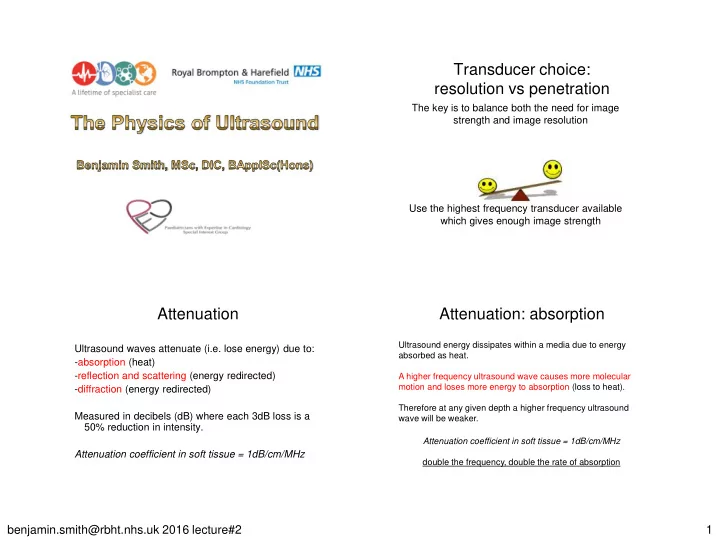

Transducer choice: resolution vs penetration The key is to balance both the need for image strength and image resolution Use the highest frequency transducer available which gives enough image strength Attenuation Attenuation: absorption Ultrasound energy dissipates within a media due to energy Ultrasound waves attenuate (i.e. lose energy) due to: absorbed as heat. -absorption (heat) -reflection and scattering (energy redirected) A higher frequency ultrasound wave causes more molecular motion and loses more energy to absorption (loss to heat). -diffraction (energy redirected) Therefore at any given depth a higher frequency ultrasound Measured in decibels (dB) where each 3dB loss is a wave will be weaker. 50% reduction in intensity. Attenuation coefficient in soft tissue = 1dB/cm/MHz Attenuation coefficient in soft tissue = 1dB/cm/MHz double the frequency, double the rate of absorption benjamin.smith@rbht.nhs.uk 2016 lecture#2 1
Percentage of Ultrasound remaining vs frequency Time-gain compensation 100% 3MHz 6MHz 79% 79% 63% 50% 63% 40% Compensation “boost” 32% to returning signal Power 50% 25% 20% 16% 40% 13% Signal strength 10% Depth/time 32% 1% 0.1% 25% 0.01% Attenuation artifacts Attenuation: reflection/scattering Large (>λ) smooth surface such as cardiac wall/valve are called specular reflectors and cause ‘mirror - like’ reflection. Acoustic shadow from Acoustic enhancement higher than expected from lower than The strength of the reflected beam is related to the difference in acoustic impedance (Z).The remaining energy deeper to the reflector attenuation (e.g. deeper expected attenuation will be weaker due to energy being redirected back/away. to calcification or (e.g. deeper to a cyst prosthesis) or pericardial fluid) Small (usually <1λ) rough/irregular surfaces which reflect in multiple directions are called scatterers. Higher f lower λ increased backscatter. Attenuation assumed to be 1dB/cm/MHz benjamin.smith@rbht.nhs.uk 2016 lecture#2 2
Maximising transmission Attenuation: reflection The strength of the reflected beam is related to the matching layer difference in acoustic impedance (Z). thin layer between the piezoelectric elements and the skin Percentage reflected = [(Z 2 – Z 1 )/(Z 2 + Z 1 )] 2 x 100% “accoustic matching” reduces reflection less attenuation and more energy transmitted % reflected at an air/soft tissue Material Acoustic interface? Impedance (Z) Air 0.0004 ?? matching layer + gel Lung 0.26 % reflected at an bone/soft Additional intermediate “accoustic matching” Soft-tissue (avg) 1.63 tissue interface? ?? reduces reflection less attenuation and more energy transmitted Bone 7.8 Ultrasound and red blood cells: Reverberation artefact Rayleigh scattering Assumption: ultrasound beam is reflected only once. Very small (<<λ) reflectors which reflect Reverberation artefact occurs when echoes bounce between two ultrasound energy concentrically, e.g. RBC highly reflective interfaces resulting in depth perception errors. (7.5µm). (recall yesterday, we worked out the λ of a 6MHz signal was 0.25mm (Q9)) Amplitude of reflected signal is less in RBC’s. Attenuation is less in RBC’s. Spontaneous contrast due to RBC aggregation. benjamin.smith@rbht.nhs.uk 2016 lecture#2 3
Refraction artifact Refraction vs mirror image artifacts Assumption: ultrasound beam travels in a straight line Refraction occurs when the ultrasound beam strikes an interface at an angle and where the speed of sound is different (according to Snells Law). Results in improper placement or duplication. Mirror-like reflector = actual object, usually displayed in correct position = object duplication as displayed The Doppler Effect The Doppler Equation (!!!) Blood flow When ultrasound interacts with a moving object Transmitted velocity (m/s) frequency (Hz) (i.e. RBC’s) the reflected frequency changes. Incident angle ± Δ f = 2 f t V cosθ If the RBC’s are traveling towards the transducer Doppler Shift c (Hz) the ultrasound wave is ”squashed” ↓λ and ↑ f Assumed speed of positive Doppler shift, i.e. red or above the line sound in soft tissue (i.e. 1540m/s) If RBC’s are traveling away: “stretched” ↑λ and ↓ f benjamin.smith@rbht.nhs.uk 2016 lecture#2 4
The Doppler Equation The Nyquist Limit (Aliasing) Assumed speed of The maximum Doppler shift ( Δ f max ) able to Doppler Shift sound in soft tissue (Hz) (i.e. 1540m/s) be displayed without aliasing. c( Δ f) V = Blood flow Determined by the sampling rate (PRF). 2 f t cosθ velocity (m/s) PRF Transmitted Incident angle Nyquist Limit: Δ f max = frequency (Hz) 2 c Δ f max = c Recall that: PRFmax = 2D 2x2D The Doppler Equation Determining the Nyquist Limit Assumed speed of sound in soft tissue (i.e. 1540m/s) c ( Δ f max ) c c c 2 V max = = x V max = 2 f t cosθ 2 f t cosθ 2x2D 8 f t cosθD Transmitted Depth Incident angle frequency (Hz) benjamin.smith@rbht.nhs.uk 2016 lecture#2 5
Determining the Nyquist Limit Determining the Nyquist Limit Assumed speed of Angle (θ) cos θ Angle (θ) cos θ % error % error sound in soft tissue 0 1.00 0 0 1.00 0 10 0.98 2 10 0.98 2 V max = 1540 2 V max = 1540 2 20 0.94 6 20 0.94 6 8 f t cosθD 8 f t cosθD 30 0.87 13 30 0.87 13 40 0.77 23 40 0.77 23 Incident angle 50 0.64 36 50 0.64 36 assumed to be 0 o Incident angle 60 0.50 50 60 0.50 50 cos 0 o = 1 Determining the Nyquist Limit S5-1 @ 5cm S5-1 @ 10cm Vmax 226cm/sec Vmax 141cm/sec V max = 1540 2 8 f t cosθD Transmitted Depth frequency (Hz) S5-1 @ 15cm Vmax 101cm/sec benjamin.smith@rbht.nhs.uk 2016 lecture#2 6
PW Doppler S5-1 S8-3 Vmax 226cm/sec Vmax 132cm/sec PW Doppler works by selectively listening S12-4 Vmax 93cm/sec All samples at 5cm depth Digital “cut and paste” PW Doppler High PRF On reflection the ultrasound wave consists of a spectrum (hence spectral Doppler) of frequencies Transducer sends out an which are digitally subtracted Doppler additional pulse before the frequency shifts ( f difference is in the audible range “sound”). From this complex wave, the original pulse has returned. process of fast fourier transformation separates In effect it doubles the PRF and each individual frequency and its amplitude and then plots this information on the spectral Doppler therefore doubles the Nyquist graph. limit. PW Doppler will display less velocities at any The disadvantage is that the single point in time vs. CW Doppler narrow spectral envelope exact origin of the Doppler shift When there is a large variation in velocities at any is not known. point in time the spectral envelope will be broader. Potential for range ambiguity This may indicate acceleration or that your gate artifact (“depth confusion”) size is too large. When the Nyquist Limit is exceeded then you will get aliasing which manifests itself as a ‘wrap - around’ signal. To counteract this, you either use high PRF PW or CW. benjamin.smith@rbht.nhs.uk 2016 lecture#2 7
CW Doppler Colour flow Doppler Effectively a multi-sampled PW from multiple sites (100-400) superimposed on a 2D image low FR!!! CW Doppler works by listening Each area sampled minimum of 3 times to calculate a Doppler frequency shift and all the time estimate mean velocity. Continuous transmission and Frame rate determined by: reception of ultrasound. • Sector size ↓width/depth↑FR No maximum velocity but…. • Packet size: The packet size is the No range resolution. number of pulses transmitted per line. ↓packet size ↑FR Same limitations as PW Doppler (i.e. Nyquist limit), however as it is detecting mean velocity the Nyquist limit is lower aliases earlier Colour and TDI Biological effects of ultrasound Thermal Effects of Ultrasound: amount of heat produced has to do with the intensity of Filters are used to discriminate between myocardium and the ultrasound, the time of exposure, and the specific absorption characteristics of tissue in colour imaging: the tissue. Thermal Index (TI): relative potential for temperature rise. Blood is a low amplitude Myocardium is a high scatterer (recall Rayleigh amplitude spectral Non-thermal/biological effects of Ultrasound: rapid and potentially large changes in scattering) with relatively reflector with relatively bubble size can occur cavitation, may increase temperature and pressure within quick velocities. slow velocities. the bubble and thereby cause mechanical stress on surrounding tissues, precipitate fluid microjet formation, and generate free radicals. Mechanical Index (MI): potential effects for cavitation, microstreaming and radiation force. Highest in PW Doppler . Recommendation: Minimise exposure time ALARA: As Low As Reasonably Achieveable. benjamin.smith@rbht.nhs.uk 2016 lecture#2 8
Recommend
More recommend