
SLIDE 1
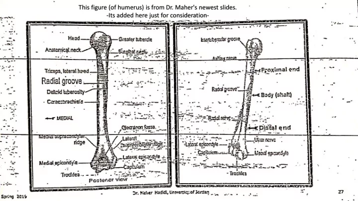
This figure (of humerus) is from Dr. Maher's newest slides.
- Its added here just for consideration-
This figure (of humerus) is from Dr. Maher's newest slides. -Its - - PowerPoint PPT Presentation
This figure (of humerus) is from Dr. Maher's newest slides. -Its added here just for consideration- Slides of Anatomy Please note : These slides are Dr. Maher Hadidi s slides of spring 2016 and were edited by the Premed Academic Team to fit
This figure (of humerus) is from Dr. Maher's newest slides.
Spring 2019
Lecture 2
Spring 2019
Voluntary 1- skeletal 700 locations :
Superficial Deep
Functions : e.g.. Body movement
Involuntary 2- Cardiac wall of heart 3- Smooth wall of organs
e.g .. Stomach
Spring 2019
1
Skeletal muscles are named according to :
radius].
Spring 2019
2
Spring 2019
Each muscle has:
Origin
Beginning.
Insertion
End.
Body (belly).
Law: When a muscle performs its action, its insertion, moves towards its origin.
3
Spring 2019
4
Spring 2019
Breast region. Contents:
1. Skin. 2. Superficial fascia. 3. Breast. 4. Deep fascia. 5. 3 Muscles:
a. Pectoralis major . b. Pectoralis minor . c. Subclavius.
5
Spring 2019
Origin: Clavicle (M ½) , Sternum and upper 6 ribs.
Insertion: humerus, lateral lip of intertubercular groove.
Nerve Supply: Medial & lateral pectoral nerves.
Action: Adducts and medially rotate the arm.
6
Spring 2019
Pectoralis Minor
O: 3rd, 4th, 5th ribs.
Ins: Coracoid process.
NS: Medial pectoral N.
Action: Depress scapula downward and forward.
7
Spring 2019
Subclavius
Origin: 1st rib.
Ins: Clavicle inferior surface.
NS: N. to Subclavius.
Action: Protects the underlying structures.
8
Spring 2019
9
PS: always refer to book for better understanding.
Spring 2019
10
A 2ply sheet of CT that
connects clavicle to the floor
Extends from both borders of
the clavicle to envelop the subclavius and pectoralis minor muscles, in order to seal the gap in between.
Pierced by:
Spring 2019
11
Spring 2019
Muscles connecting UL to vertebral col.
Arrange in layers :
12
Spring 2019
O: Clavicle, acromion and spine of scapula. Ins: Deltoid tuberosity. Action: Its fibers runs in 3 directions
Flex shoulder.
Extend shoulder joint Abduct arm 15-900 NS: Axillary Nerve.
13
Spring 2019
Supraspinatus
O: Supraspinous fossa. Ins: Greater tubercle of humerus. Action: Abducts arm 0-150. NS: Suprascapular Nerve.
Infraspinatus
O: Infraspinous fossa. Ins: Greater tubercle. Action: Lateral Rotation of arm. NS: Suprascapular N.
S i T
14
Spring 2019
Teres Minor
(L. Rounded)
O: Lat. border of Scapula. Ins: Greater tubercle of humerus Action: Lateral rotation of arm. NS: Axillary n.
Teres Major
O: Lat. border of Scapula. Ins: Medial lip of intertubercular groove Action: Adduction & Medial rotation and of arm. Remember (t.major like p.major) NS: Lower subscapular n.
15
Spring 2019
Origin: Subscapular fossa. Ins: Lesser tubercle
Action: Medial rotation
NS: Upper and lower subscapular Ns.
16
Origin: upper 8 ribs. Ins: Medial border of
scapula.
Act: Protraction of
scapula (pulls scapula forward over thoracic wall). It assists trapezius to elevate arm 1800 above shoulder .
NS: Long thoracic N.
*nerve injury cause winging of scapula
Spring 2019
17
Spring 2019
Supraspinatus Infraspinatus Teres minor Subscapularis
their insertion and blend with the fibrous capsule of the shoulder joint. They act as a handcuff that strengthen shoulder joint (superior, posterior & anterior).
18
Spring 2019
Quadrangular space
Borders:
T . minor
T . major Triceps, long head.
humerus
Contents:
.
Triangular space
Borders:
T . minor
T . major
Triceps, long head
Contents:
.
19
Spring 2019
O: Skull, spines of C1-T12 v. Ins: Its fibers runs in 3 Directions: to Clavicle.
to Acromion. to Spine of scapula.
Action:
Elevate arm. Adduct medial
Pull downward
muscle in abducting the arm 1800 above the head. NS: Spinal accessory (IX) N.
20
Spring 2019
O: T7-T12, L1- L5, Sacrum & Iliac crest. Ins: Floor of bicipital groove. Action: Extends, Adducts and Medially Rotate humerus like in
NS: Thoracodorsal N.
21