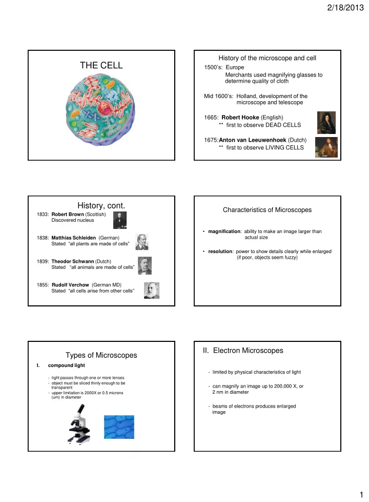
the cell
play

THE CELL 1500s: Europe Merchants used magnifying glasses to - PDF document

2/18/2013 History of the microscope and cell THE CELL 1500s: Europe Merchants used magnifying glasses to determine quality of cloth Mid 1600s: Holland, development of the microscope and telescope 1665: Robert Hooke (English) ** first


  1. 2/18/2013 History of the microscope and cell THE CELL 1500’s: Europe Merchants used magnifying glasses to determine quality of cloth Mid 1600’s: Holland, development of the microscope and telescope 1665: Robert Hooke (English) ** first to observe DEAD CELLS 1675: Anton van Leeuwenhoek (Dutch) ** first to observe LIVING CELLS History, cont. Characteristics of Microscopes 1833: Robert Brown (Scottish) Discovered nucleus • magnification : ability to make an image larger than actual size 1838: Matthias Schleiden (German) Stated “all plants are made of cells” • resolution : power to show details clearly while enlarged (if poor, objects seem fuzzy) 1839: Theodor Schwann (Dutch) Stated “all animals are made of cells” 1855: Rudolf Verchow (German MD) Stated “all cells arise from other cells” II. Electron Microscopes Types of Microscopes I. compound light - limited by physical characteristics of light - light passes through one or more lenses - object must be sliced thinly enough to be - can magnify an image up to 200,000 X, or transparent 2 nm in diameter - upper limitation is 2000X or 0.5 microns (um) in diameter - beams of electrons produces enlarged image 1

  2. 2/18/2013 Types of Electron Microscopes 2. scanning electron microscope (SEM) 1. transmission electron microscope (TEM) - three dimensional images - transmits a beam of electrons through very thinly sliced specimen - beam of electrons passed over specimens surface - dead specimens - 100,000 X mag. - 200,000 X magnification - dead specimens only - black and white only Plant Cell 3. scanning tunneling microscope CELL THEORY - computer tracks movement of probe across object - produces 3 D images - can be used on living organisms 1. all living things are composed of cells 2. cells are basic units of structure and function 3. all cells come from pre- existing cells Relationship of Surface Area to Volume How cells differ 1. size - most cells are 5-50 microns surface area ratio (limits size of cells) LENGTH OF TOTAL TOTAL SURFACE inside of cell grows faster: cubed SIDE SURFACE VOLUME AREA TO (V = L x W x H) AREA (CM2) VOLUME (CM) (CM3) RATIO outside of cell grows slower: squared 1 (1X1X6)= 6 (1X1X1)=1 6:1 (A = L x W) 24:8 2 (2X2X6)=24 (2X2X2)=8 3:1 54:27 3 (3X3X6)= 54 (3X3X3)=27 2:1 2

  3. 2/18/2013 3. internal organization - organized by nucleus and organelles 2. shape - 2 types cells - most spherical or cuboidal nerve cell 1. prokaryotes : - no nucleus or membrane - different shapes reflect bound organelles function - more primitive cells dermal epidermal cells (bacteria) white blood cells - no compartments within cytoplasm so ribosomes, enzymes, DNA free floating goblet cell - contain cell walls- made of peptidoglycans (polysaccharides connected by short chains of a. a. ) - flagella: whiplike structures for movement red blood cells Common features of all cells Types of cells, cont. - cell membrane (plasma membrane) - cytoplasm: gel like , holds cellular structures 2. eukaryotes : - cytoskeleton: microscopic protein fibers that keep cells shape - contain nucleus and membrane - ribosomes: make proteins bound organelles - DNA: controls all cell activities - unicellular eukaryotes have flagella or cilia for movement - much more complex cells CELL STRUCTURE ANIMAL CELLS Main components of eukaryotic cells Cell Membrane (plasma membrane) 1. cell membrane (outer boundary) Functions: - separates cells from surroundings 2. nucleus (control center) - regulates substance movement (bring in nutrients, remove wastes) - selectively permeable 3. cytoplasm (material between nucleus - protection and support - gives shape and flexibility and membrane) - signal receptor from other cells 3

  4. 2/18/2013 Membrane Proteins Fluid mosaic model of Cell Membrane liquid phospholipid bilayer (water insoluble, no movement through) 1. transport proteins : - involved in facilitated diffusion - polar outside (head- phospholipid, hydrophilic) (water soluble mols. not soluble in fat portion of membrane, so need help) - nonpolar inside (tail- two fatty acids, hydrophobic) - go through the entire membrane - polar outside (head- phospholipid, hydrophilic) 2 types channel : gated pores move water and ions freely in and out of cell carrier : substrate binds to site on protein protein changes shape and exposes substrate to cytoplasm Polar: hydrophilic; attracts other polar and ionic compounds Non-polar: hydrophobic; will not allow most polar molecules, ions, or large molecules to pass across the membrane animation – permeability animation – fluid mosaic 2. recognition proteins : recognizes substances (like tips of icebergs emerging from ocean surface) 3. receptor proteins : binds to specific molecules - Contain carbohydrate antennas (glycoproteins) (neurotransmitters, blood antigens, hormones) - initiates cell response - Used as chemical ID markers to differential cell types ***cell/cell recognition (immune response) ***embryo cells  tissues  organ systems 4 . enzymes : embedded in cell membrane to catalyze biochemical Nucleus reactions in cell - control center of cell: directs all cell activities - contains chromosomes - site of DNA and RNA synthesis - located in center of most cells called FLUID MOSAIC because: lipids and protein are liquid in nature and can move around each other membrane is DYNAMIC (always changing) 4

  5. 2/18/2013 Cytoplasm Structure: ( between membrane and nucleus) - contains cytosol - nuclear matrix - protein skeleton helps maintain nucleus shape – gel like material between nucleus and cell membrane - nuclear envelope (double membrane) – contains water, salts, - contains chromatin : combination of organic molecules strands of DNA and protein – in constant motion (cytoplasmic streaming) animation - nuclear pores: control substance movement amoeba animation – holds organelles - nucleoplasm : dense, protein rich Cell virtual tour - nucleolus : partially assembles ribosomes for protein synthesis Organelles Mitochondria • powerhouse of cell (cell respiration) Organelle: tiny structure that performs • provides energy for cell in form of ATP • membrane bound special functions in the cell • most numerous in cells which use a lot of energy (muscle) • self replicating, contain their own DNA to maintain life - cristae : greatly enlarge surface area of inner membrane (more area for chemical reactions of respiration) Endoplasmic reticulum: (ER) Ribosomes intercellular highway complex membrane system of folded sacs and tunnels • spherical structures which make proteins Rough ER Smooth ER • not surrounded by membrane - ribosomes stuck to membrane - no ribosomes surface - also stores and acts as an • composed of protein and nucleic acids - newly produced proteins are intercellular highway for proteins and inserted into ER enzymes • site of protein synthesis - involved in: - can be stored or exported to smooth ER - synthesis of steroids in gland - prominent in cells that make a lot - cell regulation of Ca levels of protein in muscle - cells break down toxic substances in liver cells 5

  6. 2/18/2013 Golgi Apparatus Steps of Protein Production and Transport 1. ribosomes make proteins on the rough ER- packaged into vesicles • flattened system of membranes and sacs piles on each other (like pancakes) 2. vesicles transport the newly made proteins from the rough to the Golgi apparatus • very close to ER • 3. in Golgi, proteins are processed and then packaged into NEW vesicles processes, packages, and secretes proteins to other parts of cell 4. vesicles move thru Golgi to cell membrane and release contents outside cell animation 2 Lysosomes Peroxisomes • small round vesicles that contain digestive enzymes • formed from Golgi Apparatus • contain different oxidative enzymes than lysosomes • digest and remove waste from cell (old organelles, byproducts, bact., viruses) • break down toxic substances into H 2 O 2 - detox alcohol and drugs - break down fatty acids • formed from proteins in cytosol, not Golgi animation Cytoskeleton (cell framework) Cytoskeleton Structure • maintains shape and size of cell • composed of network of long protein strands located in cytosol • not surrounded by membranes • provides movement for organelles within cytosol A. Intermediate Filaments - protein fibers coiled into cables - anchor nucleus and maintain its shape 6

Download Presentation
Download Policy: The content available on the website is offered to you 'AS IS' for your personal information and use only. It cannot be commercialized, licensed, or distributed on other websites without prior consent from the author. To download a presentation, simply click this link. If you encounter any difficulties during the download process, it's possible that the publisher has removed the file from their server.

Recommend


More recommend