
SLIDE 1
Th The I Integumentary Sy System The Skin and the Hypodermis - - PowerPoint PPT Presentation
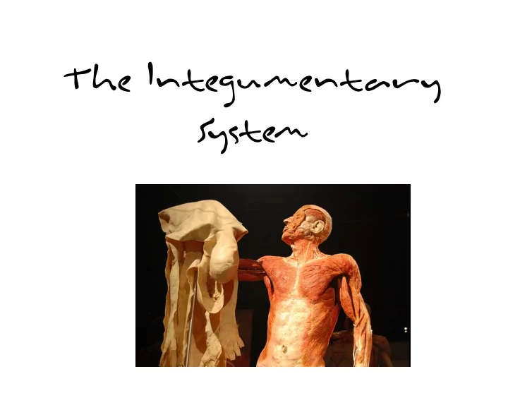
Th The I Integumentary Sy System The Skin and the Hypodermis Skin our largest organ Accounts for 7% of body weight Divided into two distinct layers Epidermis Dermis Hypodermis lies deep to the dermis 2 Skin
– Accounts for 7% of body weight – Divided into two distinct layers
– Hypodermis – lies deep to the dermis
2
3
– Cushions and insulates deeper organs – Protects body from bumps, scrapes, and cuts – Protects body from chemicals, heat, and cold – Acts as a mini‐excretory system – Screens out UV rays from the sun – Contains sensory receptors associated with nerve endings
4
– Keratinocytes
– Melanocytes – produce melanin – Merkel cells – sensory – Langerhans cells – defense cells
5
– Deepest layer of epidermis – Attached to underlying dermis – Cells actively divide – Stratum basale contains
– "Spiny" appearance caused by artifacts of histological preparation – Contains thick bundles of intermediate filaments (tonofilaments) – Contains star‐shaped Langerhans cells
6
– Consists of keratinocytes and tonofilaments
– Keratohyaline granules – help form keratin – Lamellated granules – contain a waterproofing glycolipid
– Occurs only in thick skin – Composed of a few rows of flat, dead keratinocytes
7
– Thick layer of dead keratinocytes and thickened plasma membranes – Protects skin against abrasion and penetration
8
9
tissue
vessels and nerves
– Papillary layer – includes dermal papillae – Reticular layer – deeper layer – 80% of thickness of dermis
10
– Pain & thermoregulation
11
12
– Melanin – most important pigment – made from tyrosine – Carotene – yellowish pigment from carrots and tomatoes – Hemoglobin – Caucasian skin contains little melanin
13
– Flexible strand of dead, keratinized cells – Hard keratin – tough and durable – Chief parts of a hair
14
– Medulla – central core – Cortex – surrounds medulla – Cuticle – outermost layer
15
– Hair bulb – deep, expanded end of the hair follicle – Root plexus – knot of sensory nerves around hair bulb
16
– Epithelial root sheath
epithelial root sheath
– Connective tissue root sheath
– Hair stands erect when arrector pili contracts
17
– Due to aging – Male pattern baldness
18
– Simple alveolar glands – Holocrine secretion – entire cell breaks up to form secretion
– Collects dirt; softens and lubricates hair and skin
19
20
Figure 5.1
– 99% water with some salts – Contains traces of metabolic wastes
21
– Eccrine gland
– Apocrine gland
secrete in an aprocrine fashion – rather in an eccrine or merocrine fashion as do the eccrine glands… the name has remained to avoid confusion of the two varieties of sweat glands!
22
– Made of hard keratin – Parts of the nail
– cuticle
23
– First degree burn – only epidermis is damaged – Second degree burn – upper part of dermis is also damaged
– Third degree burn – consume thickness of skin
24
– least malignant and most common – appears as a round lump
– red, pale or pearly in color – grows slowly, usually on the head, neck and upper torso – untreated can cause disfiguration
25
basal cell carcinoma extensive ulcerating basal cell carcinoma
– less common, but more dangerous than basal cell carcinoma – not as dangerous as melanoma – appears as a thickened, red, scaly spot that may bleed easily, crust or ulcerate – appears on skin most often exposed to the sun – grows over weeks to months and may spread to
26
– The most dangerous type of skin cancer – The ABCD'S of Melanoma
half.
notched or blurred.
Shades of tan, brown and black are present. Dashes of red, white and blue add to the mottled appearance.
the size of a pencil eraser). Any growth of a mole should be of concern.
– Men
shoulders & hips
– Women
27
28
– Skin thins and becomes less elastic – Shows harmful effects of environmental damage – Skin inflammations become more common
29