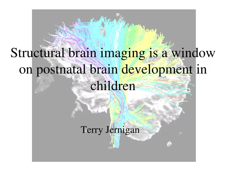
SLIDE 1 Structural brain imaging is a window
- n postnatal brain development in
children
Terry Jernigan
SLIDE 2 Old (but still widely held) View of Human Brain Structure
- All neurons are present at birth.
- Myelination of fibers occurs rapidly over the first few
years of life.
- Brain structure is “adult” at approximately age 5 (i.e.,
growth is essentially complete).
- Brain morphology is stable during late childhood,
adolescence and adulthood.
- Regressive changes of old age begin after 60.
SLIDE 3 “Brain structure is adult at approximately age 5.”
Structural MRI of young child from NIH Brain Development Study
SLIDE 4 Image Analysis for Brain Morphometry
- Stripping (Isolation of Brain
Areas)
- Bias Correction to Reduce
Signal Inhomogeneity
- Tissue Segmentation
- Anatomical Segmentation
(Within-Tissue Segmentation)
SLIDE 5
Brain Morphometry
SLIDE 6 Predictions Based on Conventional Views of Brain Morphology
- Adult brain structure in school-aged children.
- Stable brain morphological characteristics across
childhood, adolescence, and adult years.
- Atrophy of some brain structures in old age.
SLIDE 7 Age-Related Alterations of Normalized Cerebral Gray Matter Volume
.4 .45 .5 .55 .6 .65 .7 .75 .8 20 40 60 80 100
SLIDE 8
Mapping of Cortical Thinning with Longitudinal MRI Data
Gogtay et al., PNAS, 2004
SLIDE 9 Longitudinal Mapping of Cortical Thickness and Brain Growth in Normal Children
(Sowell et al.,J. Neurosci., 2004)
Widespread cortical thinning, and focal areas of cortical thickening observed longitudinally in children over 2 years, from 7 to 9.
SLIDE 10 Changes in Brain Structure in Maturing Young People
Childhood to Adolescence
(Sowell et al, NeuroImage, 1999)
Adolescence to Adulthood
(Sowell et al, Nature Neuroscience, 1999)
SLIDE 11 Age-Associated Alterations of Volumes of Subcortical Nuclei
1 2 3
Thalamus
10 20 30 40 50 60 70 80 90 100
Age
1 2 3
10 20 30 40 50 60 70 80 90 100
Age
Thalamus N.Accumbens
SLIDE 12 Curvilinear Age-Function for Hippocampal Volume
(Jernigan & Gamst, 2005)
1 2
Hippocampus
10 20 30 40 50 60 70 80 90 100
Age
Hippocampus
from N. Raz - Hedden & Gabrieli, 2005.
.12 .14 .16 .18 .2 .22 .24 .26 .28 .3 20 40 60 80 100
SLIDE 13
Why don’t young brains appear atrophied?
SLIDE 14 White Matter Growth Associated with Post-natal Proliferation of Oligodendrocytes and Myelin Deposition
1 2
Cerebral WM
10 20 30 40 50 60 70 80 90 100
Age
SLIDE 15 Summary
- During the first 2-3 decades of life, age-related tissue alterations,
presumably related to brain maturation, can be observed with morphometry.
- Though the first evidence came in the form of apparent changes in the
morphology of gray matter structures, it was suspected that much of the change was directly, or indirectly, related to continuing myelination and fiber tract development.
- However, until recently, further investigation of fiber tract maturation was
limited by the lack of sensitivity to white matter structure with existing MR methods.
SLIDE 16 Diffusion Tensor Imaging
- Measures diffusion (motion) of protons in water molecules.
- Direction of proton motion within a voxel can be described by a
“tensor”.
- Proton diffusion tends to be relatively isotropic in gray matter.
- The linear structure of fiber tracts constrains proton diffusion and
produces anisotropy.
SLIDE 17 White Matter Diffusion Properties
Apparent Diffusion Coefficient Tensor size Fractional Anisotropy Tensor shape
Low 1 Highly directional diffusion Isotropic diffusion
FA
White and Gray matter High Cerebrospinal fluid
Mean diffusivity
MD
Slide borrowed from Guido Gerig
SLIDE 18
Post-Natal Myelination is Well Visualized on MRI : Myelinating Fiber Tracts from 3 to 12 Months
SLIDE 19 Continued Fiber Tract Development Observable with DTI
(from Hermoye et al., 2006)
SLIDE 20
Lebel et al., 2007 ISMRM Meeting
SLIDE 21
Lebel et al., 2007 ISMRM Meeting
SLIDE 22
Lebel et al., 2007 ISMRM Meeting
SLIDE 23
Lebel et al., 2007 ISMRM Meeting
SLIDE 24 Diffusion Tensors and Development
- Tensor size reflects magnitude of diffusion.
– Tensors for voxels in CSF spaces are large and spherical (or isotropic): all 3 eigenvalues the same and all high. – Tensors in gray matter are smaller (less free water) but also isotropic: all 3 eigenvalues the same and all low.
- Tensor shape reflects directionality of diffusion.
– Tensors for voxels in fiber tracts are elongated (or anisotropic) presumably because diffusion of water molecules is higher within axons and along the axonal and myelin surfaces than perpendicular to the fiber tracts: principal eigenvalue (parallel diffusivity) higher than others (perpendicular diffusivity) – high “fractional anisotropy”.
- As fiber tracts mature, axons and their myelin sheaths become
larger and the water in extra-axonal space decreases.
– Less free water reduces all 3 eigenvalues (as in A) – But because axoplasmic flow and diffusion along fiber membranes is preserved or increased, principal eigenvalue (parallel diffusivity) is decreased less than other eigenvalues (perpendicular diffusivity). – Therefore, perpendicular diffusivity and fractional anisotropy are most affected by fiber tract development.
- Alterations of fiber organization (coherence, tortuosity) may
also contribute to anisotropy. A B C
SLIDE 25
“Absolute eigenvalue diffusion tensor analysis for human brain maturation”
(Suzuki et al., NMR in Biomedicine, 2003)
SLIDE 26 Contrast Between Age-Related Reductions in Parallel and Perpendicular Diffusivity
(Lebel et al., 2008)
SLIDE 27 Summary
- Although the changes may be visually subtle,
when examined closely, the brain exhibits a complex pattern of age-associated tissue alterations well into adulthood.
- We are just beginning to understand the biology
and the role that these dynamic changes play in evolving mental functions.
SLIDE 28
Relationships to Behavior
SLIDE 29 What is the significance of individual difference variability?
.2 .21 .22 .23 .24 .25 .26 .27 .28 .29 .3 Frontal Lobe Gray Matter 5 10 15 20 25 30 35 Age At Scan
Sowell, Delis, Stiles & Jernigan, 2001
Better memory retrieval was correlated with thinner (more mature) frontal cortex.
SLIDE 30 Normal Developmental Changes in Inferior Frontal Gray Matter Are Associated with Improvement in Phonological Processing: A Longitudinal MRI Analysis
(Lu et al., Cerebral Cortex, 2007)
Thickening inferior frontal cortex and thinning dorsal prefrontal cortex exhibit distinct functional correlates in the same children across the age range from 7-9.
SLIDE 31 Microstructural Correlates of Infant Functional Development: Example of the Visual Pathways
(Dubois et al., J. Neurosci, 2008)
Latency of the P1 component of the Visual Evoked Potential correlated with FA in the
independent of chronological age, in 5 - 17 week old infants.
SLIDE 32 Imaging Brain Connectivity in Children with Diverse Reading Ability
(Beaulieu et al., 2005)
32 children 8 - 12 years of age Standardized Word ID Scores 72 - 129
SLIDE 33 Tractography used to identify fiber tracts involved in developing reading fluency
(from Beaulieu et al, 2005)
SLIDE 34 Maturation of White Matter is Associated with the Development of Cognitive Functions during Childhood
(Nagy et al., 2004)
Voxel clusters in which FA correlated with spatial working memory (A-C) or reading speed (D) in 23 children aged 7 to 19. A D C B
SLIDE 35 R2 = 0.406
0.25 0.5 70 100 130 0.25 0.5 70 100 130
0.25 0.5 70 100 130
y = 0.0026x + 0.1 R
2 = 0.4199
0.25 0.5 70 100 130
Standardized Word ID Standardized Digit Recall
“Double Dissociation” in Correlation Patterns
Niogi, S. & McCandliss, B.D.(2006) Neuropsychologia Left SCR Bilateral ACR
SLIDE 36
Study of Response Inhibition in 65 7-13 year old children
SLIDE 37 Inhibitory Function Measured Using Stop Signal Task
- Principal measure of inhibitory function is the
stop signal reaction time (SSRT), which is an estimate of the time needed to inhibit, or cancel, a prepotent motor response.
- fMRI, lesion studies, and animal studies
suggest that right hemisphere inferior frontal and premotor areas are involved in this function.
SLIDE 38 Computational Morphometry: Tract-Based Spatial Statistics
(Smith et al., NeuroImage, 2006)
SLIDE 39 FA in Right IFG and Right pre-SMA account for variability in SSRT
- Individual differences in children’s inhibitory
function is related to FA differences within the neural circuit previously implicated in SST performance.
SLIDE 40 Summary
- In typically developing children and adolescents,
performance variability on behavioral tasks has been linked to morphological variability in structures within relevant neural systems, and to variability in the microstructure of fiber tracts within these systems.
- Often, the associations with performance have
been shown to remain after controlling for age.
SLIDE 41 How do we interpret these associations?
Do the relationships in children reflect the effects of variability in fiber tract development? What roles do intrinsic (genetic) factors play relative to extrinsic factors (experience)? Do experiential effects on morphology and fiber tract microstructure vary in magnitude, persistence, or functional impact as a function of age (i.e., across development)?
SLIDE 42
Cortical morphology and fiber structure exhibit relationships with performance variability in adults as well as children
SLIDE 43 Genes Exert Strong Effects on Brain Morphology
- Twin studies have revealed high heritability of
brain size, brain shape, and cortical volume.
- Genetic effects on fiber microstructure are
likely but not well characterized.
- Genetic effects on developmental trajectories
have not yet been examined.
SLIDE 44
There is growing evidence for experience related alteration of brain structure in cortex and fiber tracts
SLIDE 45 Summary
- Genetic factors undoubtedly have a strong influence
- n neural architectures -
- however, brain structure seems to exhibit subtle
alterations associated with maturation and brain aging, and with dynamic neurobiological responses to experience and training.
SLIDE 46
- MRI has contributed to an emerging picture of
protracted brain maturation and long-lasting neuroplasticity, persisting into adulthood.
- Given this, traditional structural MRI is giving
way to a new approach to imaging neurobiological as well as behavioral aspects
- f the dynamic processes involved in
development.
SLIDE 47
I.CAN
Institute for Child and Adolescent Neuroscience
SLIDE 48 Developmental Studies of Human Behavioral Phenotypic Variability
- Neural function at any point in time reflects
genetic and epigenetic factors, and the influence of both recent and remote extrinsic factors.
- Studies will focus on the evolution of
behavioral differences during development, the roles of genes and experience, and the degree to which behavioral phenotypes can be shaped by interventions.
SLIDE 49
Thank You