
SLIDE 1
Stephen Adams, Ying Buechler, Susan Taylor
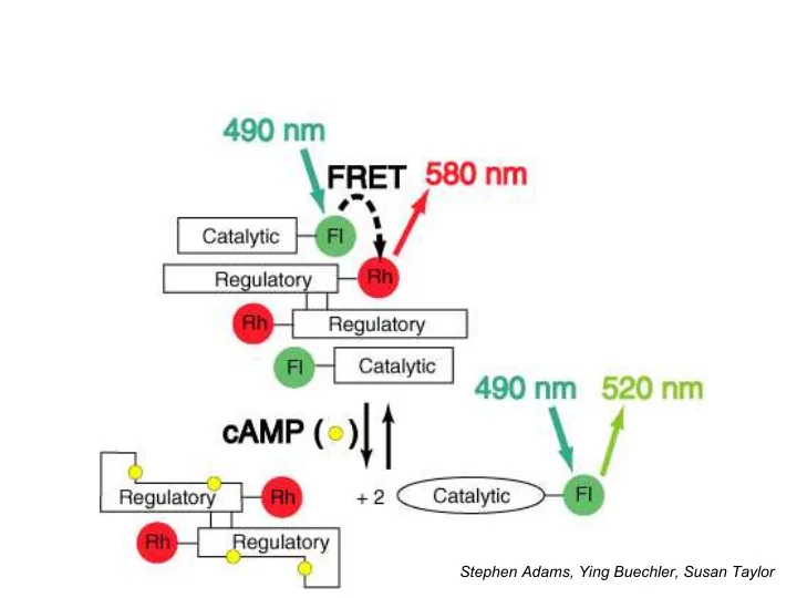
Stephen Adams, Ying Buechler, Susan Taylor Problems with cAMP sensor - - PowerPoint PPT Presentation
Stephen Adams, Ying Buechler, Susan Taylor Problems with cAMP sensor Original cAMP sensor required: expression and purification of R and C subunits of PKA in E. coli ; labeling with rhodamine and fluorescein in vitro without
Stephen Adams, Ying Buechler, Susan Taylor
– expression and purification of R and C subunits of PKA in E. coli; – labeling with rhodamine and fluorescein in vitro without destroying protein function; – reconstitution of holoenzyme – microinjection into living cells
genetically designated proteins in living cells
– Fuse naturally fluorescent proteins (ideally 2 colors), or – Devise a motif unique enough to trap small membrane- permeant dye molecules
phycobiliproteins ca. 1989? Phycocyanobilin lyase required
Photo courtesy of Claudia Mills, Friday Harbor Laboratory
GFP chromophore formation and its analogy to Asn-Gly hydrolysis
t ~ 2 hr Gly67 Tyr66 Ser65 cyclization
Proposed biosynthesis of GFP fluorophore
O2
cyclization OH- Asp Gly isoAsp Gly Asn Gly
Hydrolysis of Asn-Gly sequences
N O O H N OH N O H H OH N O O H N OH N O H
A
N O O H N OH N O H N O O H N OH O N H O H H N H O O O N H O O N H O O NH2 N H O O N H O O N O OH NH2 N H O O N O O N H O O N H O O O
Note that one molecule of H2O2 is generated for each molecule of GFP Newer work suggests that oxidation might precede dehydration (controversial)
nm), minor excitation at ~475 nm
– Broad exc. spectrum prevents usage as FRET acceptor – Ratio between two exc. peaks depends on protein concentration and past illumination
temp.
Mutations of Ser65 improve excitation spectra
WT S65 S65T
Roger Heim, Andrew Cubitt
S65A
N O N OH N H O O
S65C
BFP (Y66H…) CFP (Y66W…) Brighter GFP (S65T) YFP (T203Y…)
Examples of genetically encoded FRET sensors
Protease disrupts FRET (R. Heim) Ca2+ increases FRET (A. Miyawaki) Phosphorylation increases or decreases FRET (J. Zhang, A. Ting) cAMP disrupts FRET (M. Zaccolo, T. Pozzan (Padova))
Transgenic zebrafish embryo expressing yellow cameleon 3.60 Single confocal z-plane, imaged every 5 sec (‘mpf” = minutes post fertilization) Hide Mizuno & Atsushi Miyawaki, RIKEN
Cytosolic Ca2+ waves trigger contraction at cleavage furrows during embryonic development
Phosphorylation-dependent emission ratio of EGFR reporter, overlaid on DIC image
EGF added; FRET increases EGF washed out; FRET decreases Image taken every 5 sec; Collected over 20 min
Alice Ting
GFP-tagged HIV can be transmitted by cell-cell contact
Predominant Mode of Human Immunodeficiency Virus Transfer between T Cells Is Mediated by Sustained Env-Dependent Neutralization-Resistant Virological Synapses. Ping Chen, Wolfgang Huebner, Matthew A. Spinelli, and Benjamin K. Chen.
A High-Throughput Screen for Compounds That Inhibit Aggregation of the Alzheimer’s Peptide
Figure 1. Fluorescence-based screen using the Aβ42–GFP fusion. In the absence of inhibition, the Aβ42 portion of the fusion aggregates rapidly and causes the entire Aβ42–GFP fusion to misfold and aggregate (left). Therefore, no fluorescence is observed. However, inhibition of Aβ42 aggregation enables GFP to form its native green fluorescent structure (right). (The green part of the ribbon diagram shows the structure of GFP; the yellow part is merely a schematic representation of a nonaggregated form of Aβ42.) The triazine scaffold is shown at the center of the figure. Combinatorial diversity was introduced at sites marked X, Y, and
the figure. Compounds were added to each well, followed by E. coli cells expressing the Aβ42–GFP fusion.
Kim Woojin, Kim Yunkyoung, Min Jaeki, Kim Dong Jin, Chang Young-Tae* and Michael H. Hecht (2006) ACS Chem. Biol. 1: 461–469
Discosoma Clavularia Pocillopora Discosoma Discosoma Discosoma Discosoma Zoanthus Parazoanthus
Many tropical corals contain fluorescent proteins
First discovered by Lukyanov lab: Matz et al (1999) Nature Biotech. 17: 969-973
Structure detd. by Larry Gross, drawn by Varda Lev-Ram & Geoff Baird
The 2004 palette of nonoligomerizing fluorescent proteins
GFP-derived mRFP1-derived
605 590 nm
636 648 nm EBFP ECFP EGFP YFP (Citrine) mHoneydew mBanana mOrange tdTomato mTangerine mStrawberry mCherry mGrape1 mRaspberry mGrape2 mPlum
Evolved by SHM
Nathan Shaner et al (2004) Nature Biotech. 22: 1567-1572 Lei Wang et al (2004) Proc. Natl. Acad. Sci. USA 101: 16745-16749
Green = in mitosis Red = interphase
Cell cycle indicator using YFP and mCherry
Asako Sawano & Atsushi Miyawaki, RIKEN
BioBridge Network Meeting
Fluorescent proteins are also good educational tools in the high school classroom
Jeremy Babendure
→ Develop small peptides (≤ 12 aa) that selectively bind small synthetic molecules
through mammalian tissue
→ Develop FPs with 600-700 nm excitation
e.g. magnetic resonance
many other species
→ Develop synthetic probes localizing a variety of contrast agents at sites of high proteolytic activity
(More detail @ 4:15 PM lecture 12 Dec. 2008, G-salen, Arrhenius Laboratory, Stockholm Univ.)
A) 14 residues surrounding the biliverdin n DrCBD (PDB ID: 1ztu) were divided nto 7 groups (shown in different colors) nd targeted for mutagenesis. (B) Normalized excitation (blue) and emission
Infrared fluorescent protein based on biliverdin-binding bacterial phytochrome improves in vivo imaging
IFP1.1 + BV mKate
brightened 5 fold rel. to IFP
GFP
Adenovirally transfected livers in intact mice
Xiaokun Shu, Antoine Royant, Michael Lin, Todd Aguilera
Deinococcus radiodurans phytochrome residues targeted for mutation
BV IFP1.4 Exc. & em. spectra
ACPP colocalizes with GFP-transfected Hep2 xenografts: high magnification, after removal of skin
GFP
Quyen Nguyen & Anticancer, Inc.; Tao Jiang (Suc)e8-XPLGLAG-r9-c(Cy5)
Cy5 brightfield
(Suc)e8-Xplglag-r9-c(Cy5) d-amino acid control:
macro) is fun chemistry and can have a significant impact on cell biology and neurobiology
integrated
can make basic progress in 0.5-5 yrs (huge teams not required)
visualized in live cells
patterning are all-important
Sunset with green flash as viewed from a California lab Early work on GFP:
Douglas Prasher & Virginia Eckenrode (WHOI), Roger Heim, Andrew Cubitt.
cAMP imaging:
Stephen Adams, Susan Taylor (UCSD), Tullio Pozzan (Padova), Jin Zhang
Other CFP/YFP FRET sensors:
Atsushi Miyawaki, Varda Lev-Ram, Alice Ting
RFPs and IFPs:
Geoffrey Baird, Larry Gross, Robert Campbell, Nathan Shaner, Lei Wang, Xiaokun Shu