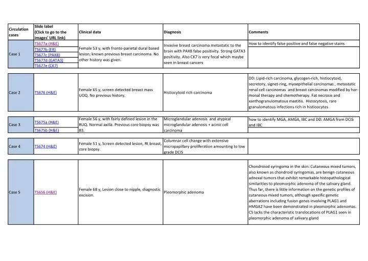

Slide label Circulation (Click to go to the Clinical data Diagnosis Comments cases images' URL link) TS677a (H&E) How to identify false positive and false negative stains Invasive breast carcinoma metastatic to the Female 53 y, with fronto-parietal dural based TS677b (ER) brain with PAX8 false positivity. Strong GATA3 Case 1 lesion; known previous breast carcinoma. No TS677c (PAX8) positivity. Also CK7 is very focal which maybe other history was given. TS677d (GATA3) seen in breast cancers TS677e (CK7) DD: Lipid-rich carcinoma, glycogen-rich, histiocytoid, secretory, signet-ring, myoepithelial carcinomas , metastatic Female 65 y, screen detected breast mass renal cell carcinomas and breast carcinomas modified by hor- Case 2 TS676 (H&E) Histiocytoid rich carcinoma UOQ. No previous history. monal therapy and chemotherapy. Fat necrosis and xanthogranulomatous mastitis. Histocytosis, rare granulomatous infections rich in histioocytes Female 56 y, with fairly defined lesion in the Microglandular adenosis and atypical how to identify MGA, AMGA, IBC and DD: AMGA from DCIS TS675a (H&E) Case 3 RUQ. Normal axilla. Previous core biopsy was microglandular adenosis + acinic cell and IBC TS675b (H&E) B3. carcinoma Columnar cell change with extensive Female 51 y, Screen detected lesion, Rt breast, Case 4 TS674 (H&E) micropapillary proliferation amounting to low core biopsy. grade DCIS Chondroiod syringoma in the skin: Cutaneous mixed tumors, also known as chondroid syringomas, are benign cutaneous adnexal tumors that exhibit remarkable histopathological similarities to pleomorphic adenoma of the salivary gland. Female 68 y, Lesion close to nipple, diagnostic Thus far, there is little information on the genetic profiles of Case 5 TS656 (H&E) Pleomorphic adenoma excision. cutaneous mixed tumors, although specific genetic aberrations including fusion genes involving PLAG1 and HMGA2 have been demonstrated in pleomorphic adenomas. CS lacks the characteristic translocations of PLAG1 seen in pleomorphic adenoma of salivary gland
Slide label Circulation (Click to go to the Clinical data Diagnosis Comments cases images' URL link) Female 57 y, diagnostic excision of superficial Case 6 TS678 (H&E) breast mass. Previous core biopsy B3. (lesion Cutaneous eccrine spiradenoma represented on the slide) Case 7 TS659 (H&E) Female 52 y.o. with UOQ mass. Invasive adenoid cystic carcinoma Female 55 y.o.; suspicious stellate lesion of the Mix of DCIS and LCIS in an area mimicking Case 8 TS660 (H&E) breast with calcifications. CSL/RS. TS663a (H&E) Female 45 y, core biopsy of suspicious mass, Case 9 TS663b (S100) Low grade secretory carcinoma. left breast. ER, PR, SMA and SMM negative. TS663c (P63) Female 67 y, wide local excison of breast mass Case 10 TS664 (H&E) in the left LIQ. Triple negative, SMA, SMM Clear cell glygogen-rich carcinoma. GCDFP-15 ngative. Malignant phyllodes tumour with osseous TS665a (H&E) heterologous differentiation, (DD metaplastic Case 11 Female 54 y, lumpectomy, Large sold mass. carcinoma with osteosarcomatous TS665b (H&E) differentiation). TS666a (H&E) TS666b (Ecadherin) Atypical microglandular adenosis with invasive Case 12 Female 46 y, VAB. TS666c (P63) NST resembling pleomorphic invasive lobular. TS666d (CK5/6) TS666e (S100) TS667a (H&E) Female 58 y, wide local excision of a 35 mm Extraskeletal myxoid chondrosarcoma (vs TS667b (Cam 5.2) Case 13 TS667c (AE1/3) rounded tumour. myoepithelial carcinoma). TS667d (S100) Case 14 TS668 (H&E) Female 81 y, mastectomy. Solid variant of adenoid cystic carcinoma.
Slide label Circulation (Click to go to the Clinical data Diagnosis Comments cases images' URL link) Grade 3 metaplastic carcinoma with spindle Case 15 TS669 (H&E) Female 74 y, lumpectomy . cell and squamous (intracystic components). Case 16 TS670 (H&E) Female 70 y, duct excision. Low grade adenosquamous carcinoma. TS672a (H&E) Female 78 y, left mastectomy; previous left Solid papillary carcinoma and invasive solid Case 17 TS672b (H&E) wide local excision of DCIS 5 years before. papillary. TS672c (ER) Female 76 y.o., history of left mastectomy Post radiotherapy angiosarcoma - poorly Case 18 TS654 (H&E) with LD reconstruction and right wide local differentiated. excision; now bilateral mastectomy. Case 19 TS679 (H&E) Female 26 y.o., nipple biopsy. Syringomatous tumour of the nipple. Case 20 TS680 (H&E) Female 64 y.o., clinically malignant mass. DLBCL. TS183a (H&E) Case 21 Female 46 y.o., Right breast lump. EPC. TS183b (H&E) TS511a (H&E) TS511c (ER) Case 22 TS511d (Synaptophysin) Female 54y old with left breast lump. SPC with invasive solid papillary features. TS511e (P63) TS511f (SMM)
Slide label Circulation (Click to go to the Clinical data Diagnosis Comments cases images' URL link) TS512a (H&E) TS512b (H&E) TS512c (P63) SPC showing features overlapping between in Case 23 Female 90y old with nipple discharge. TS512d (SMM) situ and invasive. TS512e (CK14) TS512f (CD10) TS514 (H&E) TS514 b (B catenin) TS514c (CK5/6) Case24 Female 40y old with right breast lump Fibromatosis. TS514d (CD34) TS514e (AE1/AE3) TS514f (CK14) TS287a (H&E) TS287b (H&E) F 80y with left breast lump. B5a core biopsy. TS287c (ER) Case 25 TS287d (SMM) Excision specimen shows two adjacent lesions Solid papillary carcinoma + Benign papilloma. TS287e (CK5/6) (representative section with IHC). TS287f (P63) TS287g (CK14)
Recommend
More recommend