
SLIDE 1
Skin injuries in interventional procedures Madan Rehani, PhD - - PowerPoint PPT Presentation
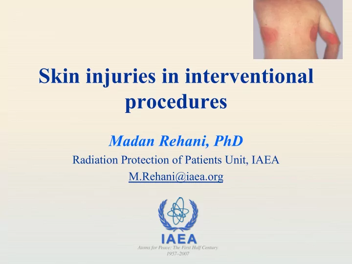
Skin injuries in interventional procedures Madan Rehani, PhD Radiation Protection of Patients Unit, IAEA M.Rehani@iaea.org Skin injury Although called skin injury severe injuries can extend upto subcutaneous fat and muscle Epidermis
2
3
4
5
6
Effect Single dose Threshold (Gy) Onset Early transient erythema 2 Hours Main Erythema 6 ~10 d Temporary hair loss 3 ~3 wk Permanent hair loss 7 ~3 wk Dry desquamation 14 ~4 wk Moist desquamation 18 ~4 wk Secondary ulceration 24 >6 wk Late erythema 15 ~6 – 10 wk Ischemic dermal necrosis 18 >10 wk Dermal atrophy (1st phase) 10 >14 wk Dermal atrophy (2nd phase) 10 >1 yr Induration (Invasive Fibrosis) 10 Telangiectasia 10 >1 yr Late dermal necrosis >12? >1 yr Skin cancer not known >5 yr
7
8
9
10
11
12
13
14
15
16
17
18
19
20
21
22
2 4 6 8 10 12 14 50 100 150 200 250 300 350 400 450 Fluoroscopy Time (min) Cumulative Dose (Gy)
Fluoroscopy Time (min) Cumulative Dose (Gy)
23
24
n 8 x 10 chip matrix n 4 cm x 4 cm grid spacing
25
From MARTIR EC training programme (pub no. 199) www.europa.eu.int/comm/environment/radprot/#news
26
RADIOCHROMIC FILMS:
dose range: 0.1-15 Gy
energy (60 - 120 keV)
image (with a flatbed scanner)
27
Example of dose distribution in a Coronary angiography procedure shown on a radiochromic film √
28
29
30
31
32
33
34
35
Lesion required grafting.
36
37
38
39
40