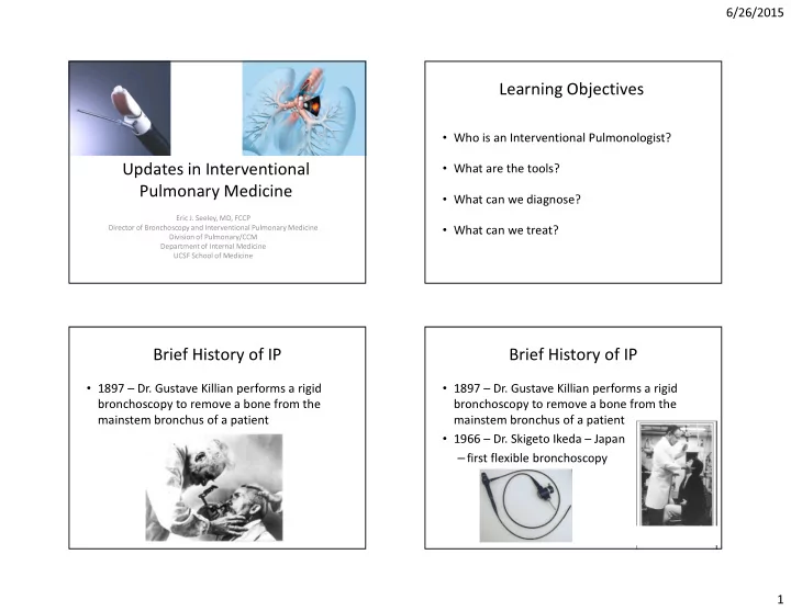

6/26/2015 Learning Objectives • Who is an Interventional Pulmonologist? • What are the tools? Updates in Interventional Pulmonary Medicine • What can we diagnose? Eric J. Seeley, MD, FCCP Director of Bronchoscopy and Interventional Pulmonary Medicine • What can we treat? Division of Pulmonary/CCM Department of Internal Medicine UCSF School of Medicine Brief History of IP Brief History of IP • 1897 – Dr. Gustave Killian performs a rigid • 1897 – Dr. Gustave Killian performs a rigid bronchoscopy to remove a bone from the bronchoscopy to remove a bone from the mainstem bronchus of a patient mainstem bronchus of a patient • 1966 – Dr. Skigeto Ikeda – Japan – first flexible bronchoscopy 1
6/26/2015 Who becomes an Interventional What does an Interventional Pulmonologist? Pulmonologist do? • It depends on their tools • Most did a residency in internal medicine • In general involved in the work-up and • Then a fellowship in Pulmonary CCM diagnosis of thoracic malignancies • And then a formal or informal fellowship in • Also involved in therapy Interventional Pulmonary Medicine – Airway Recanalization • This is a non-ACGME fellowship – Tumor Ablation – Fiducial placement • Evolving board exam, but not required • Tools offer access to: – Pleural Space, Airways, Lung Parenchyma Traditional Bronchoscopy What are the tools? Anatomic Considerations 17-25 generations Trachea 20-25 mm Mainstem 12-16 mm Segmental 5-8 mm Therapeutic scope 5.8 mm Diagnostic 5.2 mm OD 2
6/26/2015 Traditional Bronchoscopy Traditional Bronchoscopy Anatomic Considerations Anatomic Considerations 17-25 generations 17-25 generations Trachea 20-25 mm Trachea 20-25 mm Mainstem 12-16 mm Mainstem 12-16 mm Segmental 5-8 mm Segmental 5-8 mm Therapeutic scope 5.8 mm Therapeutic scope 5.8 mm Diagnostic 5.2 mm OD Diagnostic 5.2 mm OD But there is so much more…LNs But there is so much more…nodules 3
6/26/2015 Endobronchial Ultrasound (EBUS) Endobronchial Ultrasound (EBUS) But there is so much more…LNs But there is so much more…LNs 4
6/26/2015 EBUS EBUS – Image with Doppler Mediastinoscopy? EBUS – Image with Doppler What LNs are accessible? 5
6/26/2015 EBUS-Therapeutic options. Endobronchial Ultrasound Treat Diagnosis – Obtain tissue from enlarged LNs • cancer, sarcoid, lymphoma, granulomatous infections – Allows for LN staging for lung cancer – Can place fiducials for XRT – Can be performed at the same time as EMN – Come and go procedure – Can deliver Ampho to Aspergillomas – Can obtain enough tissue for molecular diagnostics EMN(B) Comparable to GPS in the lungs (electromagnetic navigation bronchoscopy) 6
6/26/2015 Procedure: navigation views Procedure: at the target 28 | 27 | Nodule-lung windows EMN- case illustration • 57 yo man of Japanese ancestry • Presented with respiratory symptoms including cough • Found to have a 1.2 cm nodule in lung • Mildly PET positive • Recommended lobectomy • Small hilar lymph nodes 7
6/26/2015 Paragonimus Westermanii Before and after treatment with EMN (electromagnetic navigation bronchoscopy) Praziquantel – Performed through ETT (fluoro vs. OR) – Can biopsy lesions almost anywhere in the lung down to 5 mm in size – Can biopsy, place fiducials, dye for localization – Easily combined with EBUS for full staging – Overlap with CT-FNA, if touching pleura or no “easy airway” would send for CT-FNA – Faster diagnosis and staging with combined EMN/EBUS 8
6/26/2015 Once procedure…comprehensive Once procedure…comprehensive diagnosis and treatment diagnosis and treatment 68 yo smoker with severe emphysema 68 yo smoker with severe emphysema High risk TTNA High risk TTNA Not a surgical candidate Not a surgical candidate 1. Tissue DX with EMN 1. Tissue DX with EMN 2. Staging with EBUS Once procedure…comprehensive Rigid Bronchoscopy diagnosis and treatment 68 yo smoker with severe emphysema High risk TTNA Not a surgical candidate 1. Tissue DX with EMN 2. Staging with EBUS 3. If EBUS is negative fiducials could be placed for XRT 9
6/26/2015 Rigid Bronchoscopy- Cryotechnologies Why would we do this? • Contact • Requires Jet Ventilation • Cryoprobe • Allows more stable • Freezes to -90 access to distal trachea • Cryogen is NO 2 or CO 2 • Allows access for larger • Adheres to everything tools • Good for: • Provides opportunity to – Tumor extraction remove large objects – Foreign body extraction (tumor, foreign body) – parenchymal lung biopsy? • Provides access for advanced airway tools Cryoprobe extraction: Case Cryoprobe extraction: Case Before Cryoprobe extraction, cryospray, bronchoplasty 10
6/26/2015 Cryotechnologies Cryoprobe extraction: Case • Non Contact – Cryospray – Usually via Rigid Bronch – Obviates need for stent – Gas expands 700 x • risk of barotrauma – Cools to -196 F – Can be combined with bronchoplasty or cryoprobe extraction of airway tumor – ECM resistant to cryo-injury due After Before to lower water content Cryoprobe extraction, cryospray, bronchoplasty Bronchial Thermoplasty for Severe Asthma Bronchial Thermoplasty (BT) • a - 3 Procedures, 3 weeks apart - Deliver Thermal Energy to airway smooth muscle - Most common side effect is asthma exacerbation - Unclear which population might benefit most Castro et al AJRCCM 2010 11
6/26/2015 Trials in IP RePneu Trial for Emphysema • PneumRx – coils for • Endobronchial Lung Volume Reduction LVRC in emphysema – Lung volume reduction coils • RCT finished – Lung volume reduction valves • Now entering cross over • Endobronchial Valves for BPF PulmonX – Lung Volume Reduction for Spiration trial for BPF (VAST) Emphysema – LIBERATE TRIAL • Compassionate use for BPF * Requires screen for colateral ventilation before insertion of valve 12
6/26/2015 Conclusions • IP allows for access to lung beyond the optical reach of a traditional bronchoscopy Questions? • Can be used for the diagnosis, staging and therapy in lung cancer • Advanced tools allow for extraction/ablation of airway tumors • New tools may provide additional options for asthma, emphysema, BPF 13
Recommend
More recommend