SLIDE 1
SLIDE 2
Reflex Arc for Stretch Reflex
SLIDE 3
Figure 13.2b, p. 419
Muscle Spindles
SLIDE 4
Proprioceptors
muscle spindle
Figure 16.9a Neuroscience, 2nd Edition (2001)
Golgi tendon organ
Figure 16.11a Neuroscience, 2nd Edition (2001)
SLIDE 5
The Stretch Reflex in Action
Figure 16.9b Neuroscience, 2nd Edition (2001)
SLIDE 6 Reciprocal Innervation in the Stretch Reflex
- muscle spindle afferent directly excites somatic motor neuron to
same muscle
- muscle spindle afferent excites an inhibitory interneuron that
prevents activation of somatic motor neurons to antagonists muscle
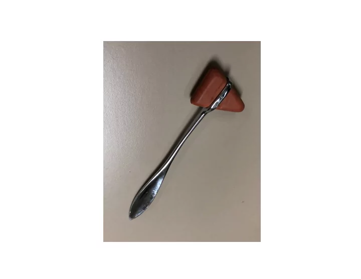
SLIDE 7
What structure is hit with the reflex hammer? What muscles are stretched? What muscle action occurs as a consequence? Why do this test?
SLIDE 8 The Stretch Reflex is a Negative Feedback Loop to Maintain Muscle Length
- loss of descending inhibition from CNS in upper motor neuron disorders
- hypertonia; hyperreflexia; clonus
- positive Babinski sign
SLIDE 9 Plantar Reflex: Babinski Sign
normal adult response positive Babinski sign
- positive Babinski sign occurs in infants and in upper motor neuron disorders
SLIDE 10 Stretch Reflexes to Test
- patellar tendon (knee jerk)
- Achilles tendon (ankle reflex)
- triceps tendon
- finger flexors
SLIDE 11 Patient B
Diagnosis
- Is it peripheral neuropathy?
- If so, what is the cause?
SLIDE 12 Sensory Nerve Conduction Study: Recording Setup
ground recording electrode stimulating electrode
To determine conduction velocity:
- measure distance along nerve between negative pole of
stimulating electrode and positive pole of recording electrode
- divide by time between stimulus and start of recorded
response
SLIDE 13 Sensory Nerve Conduction Study
stimulus artifact recorded response stimulus start of recorded response
recorded response is due to the sum of all the action potentials in axons
recording electrode
SLIDE 14
Electromyography
stimulate nerve and record from muscle What is the specific electrical activity in the muscle cells that is being measured in and EMG recording?
SLIDE 15
Motor Unit: Somatic Motor Neuron and all the Muscle Fibers it Innervates
EMG signal: summed APs of all the muscle fibers in the motor unit
SLIDE 16
Peripheral Neuropathy
demyelinating: decreased conduction velocity example: Guillain-Barré axonal: decreased amplitude (fewer axons firing action potentials) example: diabetic neuropathy polyneuropathy: multiple nerves affected; generalized disorder diabetic neuropathy: usually sensory-predominant, distal axonal polyneuropathy
SLIDE 17
Figure 36-14A in Principles of Neural Science (2000) 4th edition
Glove and Stocking Pattern
SLIDE 18 Terminology
paresthesia: abnormal sensation allodynia: pain in response to a non-painful stimulus HbA1c: percentage of glycated hemoglobin
- used to monitor glycemic control
- used to diagnose diabetes mellitus
SLIDE 19
Examination
SLIDE 20 Treatments for Diabetic Neuropathy
- glycemic control
- treat cardiovascular issues
- pain medications to control symptoms
SLIDE 21 Patient C
Possible causes of weakness:
- CNS damage (upper motor neuron disorder)
- peripheral neuropathy
- myopathy
- neuromuscular junction disorder
SLIDE 22
SLIDE 23 Ptosis
- due to weakness in skeletal muscles that retract the eyelid
SLIDE 24 Figure 12.12
Creatine Kinase: a Marker for Muscle Cells
SLIDE 25 Myasthenia Gravis
- autoimmune disorder
- antibodies to nicotinic acetylcholine receptor at NMJ
most common cause, but can be caused by other autoantibodies
- decreases the size of the muscle EPSP
- diagnosed by tests for antibodies, repetitive nerve
stimulation test
SLIDE 26
Figure 1 in Lee, I., et al “Weak and Winded” NEJM 381: 76-82
Decline in Amplitude of Muscle Response to Repetitive Nerve Stimulation
SLIDE 27
Repetitive Nerve Stimulation Test
SLIDE 28
Steps in Muscle Excitation
SLIDE 29
Muscle EPSP
SLIDE 30
Repetitive Nerve Stimulation Test
SLIDE 31 Treatment of Myasthenia Gravis
- pyridostigmine
- immunosuppressive drugs
SLIDE 32 “Diagnosing Myasthenia Gravis with an Ice Pack” NEJM (2016) 375: e39
- ice pack decreases acetylcholinesterase activity to improve symptoms
Ice Pack Test