REFERENCES The patient in this report was only 23 years with a giant - - PDF document

references the patient in this report was only 23 years
SMART_READER_LITE
LIVE PREVIEW

REFERENCES The patient in this report was only 23 years with a giant - - PDF document

CASE REPORT Bhuyan L et al.: Megasized Salivary Gland Calculi U nusual P resentation of M egasized S alivary G land C alculi: C ase R eport and R eview of L iterature Lipsa Bhuyan 1 , Sarat Nayak 2 , Suryakanti Nayak 3 , Kailash Chandra Dash 4


slide-1
SLIDE 1

International Journal of Oral Health and Medical Research | ISSN 2395-7387 | NOVEMBER-DECEMBCER 2016 | VOL 3 | ISSUE 4

69 CASE REPORT

Bhuyan L et al.: Megasized Salivary Gland Calculi

Correspondence to:

  • Dr. Lipsa Bhuyan, Senior Lecturer, Department of Oral and

Maxillofacial Pathology, Kalinga Institute of Dental Sciences,

  • KIIT. Bhubaneswar, Odisha

Contact Us: www.ijohmr.com

Unusual Presentation of Megasized Salivary Gland Calculi: Case Report and Review of Literature

Lipsa Bhuyan1, Sarat Nayak2, Suryakanti Nayak3, Kailash Chandra Dash4

Sialadenosis is inflammation of salivary gland whose etiology range from a simple infection to autoimmune cause. Salivary gland calculus constitutes of 50% of major salivary gland diseases. It seldom attains a size greater than 1.5

  • cms. When infected, it commonly causes considerable amount of pain and swelling. We hereby report a case of giant

salivary gland lith in the warton’s duct and a review of literatature on its etiopathogenesis and multifarious management modalities.

KEYWORDS: Sialadenosis, Sialolithiasis, Salivary Gland Calculi, Wharton’s duct calculi

AA

aaaasasasss

Non-neoplastic growth in submandibular area may be caused by sialadenitis, mumps, Sjögren syndrome, cysts and infections.1 Sialolith, a common disease of salivary gland, is usually associated with acute and chronic

  • infection. Submandibular gland is a host of 80-90% of

sialoliths because of higher salivary viscosity and salivary

  • stasis. 5-10% occurs in parotid gland and 1-2% in

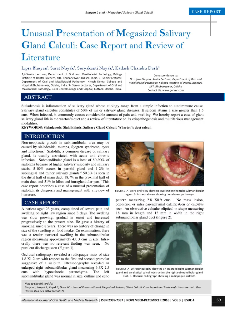
sublingual and minor salivary glands.2 50.3% is seen in the distal half of main duct, 18.7% in the proximal half of main duct and 31% in hilus and intraglandular part.3 This case report describes a case of a unusual presentation of sialolith, its diagnosis and management with a review of literature. A patient aged 23 years, complained of severe pain and swelling on right jaw region since 3 days. The swelling was slow growing, gradual in onset and increased progressively to the present size. He gave a history of smoking since 8 years. There was no history of change in size of the swelling on food intake. On examination, there was a tender extraoral swelling in the submandibular region measuring approximately 4X 3 cms in size. Intra-

  • rally there was no relevant finding was seen. No

purulent discharge seen (Figure 1). Occlusal radiograph revealed a radiopaque mass of size 1.8 X1.2 cm with respect to the first and second premolar suggestive of a sialolith. Ultrasonography revealed an enlarged right submandibular gland measuring 3.3X 2.5 cms with hypoechoeic parenchyma. The left submandibular gland was normal in size, outline and echo pattern measuring 2.8 X0.9 cms . No mass lesion, collection or intra parenchymal calcification or calculus

  • seen. An obstructive calculus eliptical in shape measuring

18 mm in length and 12 mm in width in the right submandibular gland duct (Figure 2).

How to cite this article: Bhuyan L, Nayak S, Nayak S, Dash KC. Unusual Presentation of Megasized Salivary Gland Calculi: Case Report and Review of Literature. Int J Oral Health Med Res 2016;3(4):69-71.

INTRODUCTION

1,4-Senior Lecturer, Department of Oral and Maxillofacial Pathology, Kalinga Institute of Dental Sciences, KIIT. Bhubaneswar, Odisha, India. 2- Senior Lecturer, Department of Oral and Maxillofacial Pathology, Hitech Dental College and Hospital,Bhubaneswar, Odisha, India. 3- Senior Lecturer, Department of Oral and Maxillofacial Pathology, S.C.B Dental College and Hospital, Cuttack. Odisha. India.

ABSTRACT CASE REPORT

Figure 1- A- Extra-oral view showing swelling on the right submandibular

  • region. B- Intra-oral view showing no relevant pathology.

Figure 2- A- Ultrasonography showing an enlarged right submandibular gland and an eliptical calculi obstructing the right submandibular gland

  • duct. B- Occlusal radiograph showing a radiopaque sialolith.
slide-2
SLIDE 2

International Journal of Oral Health and Medical Research | ISSN 2395-7387 | NOVEMBER-DECEMBCER 2016 | VOL 3 | ISSUE 4

70 CASE REPORT

Bhuyan L et al.: Megasized Salivary Gland Calculi

The lith could only be felt when palpated deep in the floor of the mouth. Local anesthesia was induced and a longitudinal mucosal incision was placed through the duct wall superiorly. A blunt dissection of the tissues was done and the sialolith was tracked down. It was then retrived (Figure 3). Pus discharge was observed. Saline irrigation was done. Vicryl sutures was placed to approximate the wound. Medications prescribed included antibiotics, analgesics along with sialogouges to aid removal of residual calculi. Healing was satisfactory and patient was recall for follow up. No recurrence was seen after one year of surgery. Sialolithiasis is a common disorder constituting 50% of all salivary gland pathologies with a frequency of 0.15%

  • f adult population and slightly male predilection. It

consists of 82% inorganic and 18% organic material. It may be of various shapes, sizes and texture. A usual size

  • f 5 to 10 mm is seen. A size greater than 15 mm can be

considered as a giant sialolith.2 Peak incidence is between 30 to 60 years. Multiple calculi is observed in 25% of cases and bilateral calculi in approximately 2% of cases.4 The patient in this report was only 23 years with a giant single unilateral calulus measuring 1.8 X1.2 cm. Various theories have been proposed for etiopathogenesis

  • f formation of sialolith. A relative stagnation of calcium

rich saliva can cause deposition of calcium salts around a nidus of organic component such as altered salivary mucin, bacteria and desquamated epithelial cells. An alteration in calcium phosphate solubility due to unknown metabolic phenomenon increases the concentration of salivary bicarbonate leading to calcium and phosphate ions precipitation. Another theory suggests that it can develop secondary to sialadenitis and is associated with its duration and symptoms.5 Studies have shown association between incidence of sialolithiasis and smoking.6,7,8 In the present case, the patient was a heavy smoker was could have been the risk factor herein. Sialolithiasis is usually associated with sialadenosis resulting in pain and discomfort due to obstruction of salivary secretion. A thorough examination is necessary to locate the sialolith before it takes up a massive

  • dimension. Usually submandibular gland duct liths

presents with a hard swelling in the floor of the mouth.5 In our case, the patient presented with pain and swelling but did not have any intraoral presentation. Even the history of increase in swelling on food intake was absent. Mandibular occlusal radiograph, ultrasound, computed beam tomography, sialography and scintigraphy are a few diagnostic imaging studies that can be carried out to locate the stone in salivary gland.9 We employed mandibular occlusal radiograph and ultrasonograpy to visualize the location and size of the sialolith. Treatment modalities range from conservative to surgical management depending on the size and location. Sialogogues aid in flushing out of small sized salivary

  • liths. If present on distal third of duct, it can be milked

and extracted through the duct orifice. It can also be surgically released by a minor mucosal incision in the floor of the mouth.5 Advanced method of treatments have emerged like extracorporeal shock-wave lithotripsy, laser intra-corporeal lithotripsy, interventional radiology, sialoendoscopy, the video-assisted conservative surgical removal of parotid and sub-mandibular calculi and botulinum toxin therapy.10 In the present study, the calculus was present in the proximal third of the duct and therefore surgical method was chosen. Sialolithiasis a common obstructive salivary gland disorder can often effect quality of life. Warning signs like pain, increase in swelling on food intake and sometimes foul taste. Hard swelling in the floor of the mouth can be a positive indication of salivary calculus. Calculus present intraglandularly or proximal to the duct can be difficult to locate on palpation. A thorough examination is necessary to locate the sialolith before it takes up a massive dimension.

1. Eleftheriadis I, Papadimitriou P, Tzelepi H. Submandibular swelling and its differential diagnosis. Hell Period Stomat Gnathopathoprosopike Cheir. 1990 Jun;5(2):59-68. 2. Mathew Cherian N, Vichattu SV, Thomas N, Varghese A. Wharton’s Duct Sialolith of Unusual Size: A Case Report with a Review of the Literature. Case Reports in Dentistry. 2014;2014: 373245. 3. Lustmann J, Regev E, Melamed Y. Sialolithiasis. A survey

  • n 245 patients and a review of the literature. Int J Oral

Maxillofac Surg1990;19:135-8. 4. Som PM, Brandwein MS (2003) Salivary glands: anatomy and pathology. In: Som PM, Curtin HD (eds) Head and neck imaging, 4th edn. Mosby, St. Louis, Missouri, pp 2005–2133 5. Siddiqui SJ. Sialolithiasis: an unusually large submandibular salivary stone. Br Dent J. 2002;193(2):89– 91.

Figure 3- A- Local anesthesia administered. B- Incison placed. C- sialolith located. D-The salivary gland lith

DISCUSSION CONCLUSION REFERENCES

slide-3
SLIDE 3

International Journal of Oral Health and Medical Research | ISSN 2395-7387 | NOVEMBER-DECEMBCER 2016 | VOL 3 | ISSUE 4

71 CASE REPORT

Bhuyan L et al.: Megasized Salivary Gland Calculi 6. Stanford KA. Submandibular Duct Fistula Caused by a Large Sialolith: Incidental Finding in a Barium Swallow Study.Radiol Technol. 2015 Jul-Aug;86(6):610-3. 7. Almasri M. Management

  • f

giant intraglandular submandibular sialolith with neck fistula. Ann Dent 2005;12(1):41-46. 8. Huoh KC, Eisele DW. Etiologic factors in sialolithiasis. Otolaryngol Head Neck Surg 2011;145(6):935 -939. 9. Gupta A, Rattan D, Gupta R. Giant sialoliths of submandibular gland duct: Report of two cases with unusual shape. Contemporary Clinical Dentistry. 2013;4(1):78-80.

  • 10. Capaccio P, Torretta S, Ottaviani F, Sambataro G,

Pignataro L. Modern management of obstructive salivary diseases. Acta Otorhinolaryngologica Italica. 2007;27(4):161-172.

Source of Support: Nil Conflict of Interest: Nil