
SLIDE 1
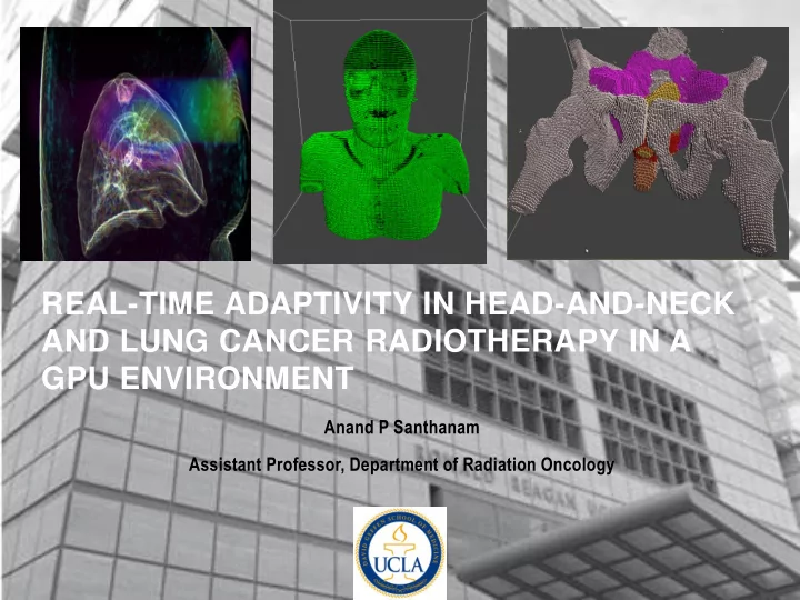
REAL-TIME ADAPTIVITY IN HEAD-AND-NECK AND LUNG CANCER RADIOTHERAPY IN A GPU ENVIRONMENT
Anand P Santhanam Assistant Professor, Department of Radiation Oncology
REAL-TIME ADAPTIVITY IN HEAD-AND-NECK AND LUNG CANCER RADIOTHERAPY - - PowerPoint PPT Presentation
REAL-TIME ADAPTIVITY IN HEAD-AND-NECK AND LUNG CANCER RADIOTHERAPY IN A GPU ENVIRONMENT Anand P Santhanam Assistant Professor, Department of Radiation Oncology OUTLINE Adaptive radiotherapy for head and neck and lung cancer Key tools
Anand P Santhanam Assistant Professor, Department of Radiation Oncology
2
3
4
5
6
7
8
Radiation Oncology*Biology*Physics. 2014
10
11
12
13
per element
15
16
17
12
19
Case 3 Case 4 Case 5 Case 1 Case 1 Case 2
22
latencies than Global memory
23 / 5
24 / 6
25 / 6
26
27