
SLIDE 1
Yale West Campus Materials Characterization Core (MCC) ywcmatsci.yale.edu
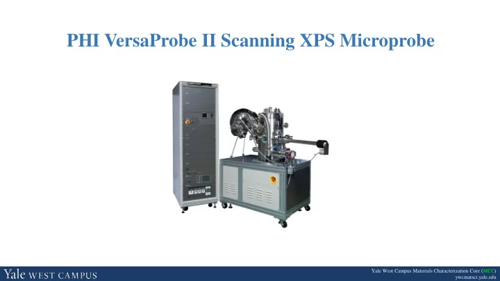
PHI VersaProbe II Scanning XPS Microprobe Yale West Campus Materials - - PowerPoint PPT Presentation
PHI VersaProbe II Scanning XPS Microprobe Yale West Campus Materials Characterization Core (MCC) ywcmatsci.yale.edu Core Policies DO NOT let other people use the facility under your account. DO NOT try to fix parts or software issues by
Yale West Campus Materials Characterization Core (MCC) ywcmatsci.yale.edu
Materials Characterization Core (MCC) ywcmatsci.yale.edu
2/20
Yale West Campus
Materials Characterization Core (MCC) ywcmatsci.yale.edu
3/20
Yale West Campus
detector electron
Vacuum or Ambient pressure
photoelectrons as the function of binding energy
Materials Characterization Core (MCC) ywcmatsci.yale.edu
4/20
Yale West Campus
detector electron
Vacuum or Ambient pressure
photoelectrons as the function of binding energy
Materials Characterization Core (MCC) ywcmatsci.yale.edu
5/20
Yale West Campus
Materials Characterization Core (MCC) ywcmatsci.yale.edu
6/20
Yale West Campus
within the sample volume
KE (measured) = hν - BE – Φspec BE = hν - KE - Φspec KE (KLL) = BE(K) – BE(L2) – BE(L3) Ionization (initial state) Relaxation and Emission (final state)
Auger Electron Φ BE L3 L1 L2 X-ray Fluorescence K UV Photoelectron Vacuum VB 2p3/2 2p 1s X-ray Photoelectron EF Φ hν 2s 2p1/2 hν e-
Materials Characterization Core (MCC) ywcmatsci.yale.edu
7/20
Yale West Campus
KE BE
Materials Characterization Core (MCC) ywcmatsci.yale.edu
8/20
Yale West Campus
l = 0 s 1 p 2 d 3 f j = l ± s, s = 1/2
Spin-orbital splitting with l > 0
Orbital l j Degeneracy (2j + 1) Peak area ratio Electron level s 1/2 1
p 1 1/2, 3/2 2, 4 1 : 2 2p1/2, 2p3/2 d 2 3/2, 5/2 4, 6 2 : 3 3d3/2, 3d5/2 f 3 5/2, 7/2 6, 8 3 : 4 4f5/2, 4f7/2
Materials Characterization Core (MCC) ywcmatsci.yale.edu
9/20
Yale West Campus
UV lamp Hemispherical analyzer X-ray source Flood gun Sample UHV chamber (low 10-7 – 5x10⁻⁸ Pa Ion gun e- e-
Ar+
Detector Lens Pumps
UHV system (< 10-8 Torr)
length Electron analyzer
energies
X-ray source
1256.6 eV
crystal Low-energy electron flood gun
Ion gun
sources may be required
Sample holder Electron energy analyzer X-ray source
PHI VersaProbe II XPS
E-neutralizer
Materials Characterization Core (MCC) ywcmatsci.yale.edu
10/20
Yale West Campus
X-ray lines Line Energy (eV) Width (eV) Mg Kα1,2 1253.6 0.70 Al Kα1,2 1486.6 0.85
K (1s) L (2s) L2 (2p1/2) L3 (2p3/2) M1 (3s) M2,3 (3p) M4,5 (3d) Kα1 Kα2 Kβ
Materials Characterization Core (MCC) ywcmatsci.yale.edu
11/20
Yale West Campus
n λ = 2dsinθ For quartz (1010) surface: n = diffraction order d = 0.42 nm (lattice constant) θ = 78.5º λ = 0.83 nm for Al Kα
Materials Characterization Core (MCC) ywcmatsci.yale.edu
12/20
Yale West Campus
Pass energy: Analyzer Resolution: V0: the median equipotential surface of radius r V: the potential applied between inner (radius b) and outer (radios a) shells w: entrance and exit slit widths 𝜀𝛽: angular deviation of the electron trajectories at the entrance with respect to the center line r = a+b 2 Where the mean radius 𝐹0 = 𝑓𝑊
0 =
𝑊 𝑐 𝑏 − 𝑏 𝑐
a b
r
𝜺𝜷 V2<0
w w
V
∆𝐹 = 𝐹0 𝑥 𝑏 + 𝑐 + 𝜀𝛽2 4 For the PHI SCA : 𝐹0 = 0.56𝑊 ∆𝐹 = 0.015𝐹0 and Typical 𝐹0 = 100 eV ∆𝐹 = 1.5 eV
Materials Characterization Core (MCC) ywcmatsci.yale.edu
13/20
Yale West Campus
http://www.eag.com/mc
for Chemical Analysis (ESCA)
Typical Analysis Depths for Techniques
XPS detects electron signals in the near surface region (0 ~ 10 nm)
Materials Characterization Core (MCC) ywcmatsci.yale.edu
14/20
Yale West Campus
http://www.eag.com/mc
reached below 10 µm
range
Materials Characterization Core (MCC) ywcmatsci.yale.edu
15/20
Yale West Campus
Materials Characterization Core (MCC) ywcmatsci.yale.edu
16/20
Yale West Campus
“Universal Curve” - λ (IMFP) vs kinetic energy λ = 1 ~ 3.5 nm for X-ray photoelectrons
inelastic collisions.