Patellofemoral Arthroplasty .align the patella!!!! Phil Davidson, - - PowerPoint PPT Presentation

patellofemoral arthroplasty align the patella
SMART_READER_LITE
LIVE PREVIEW

Patellofemoral Arthroplasty .align the patella!!!! Phil Davidson, - - PowerPoint PPT Presentation

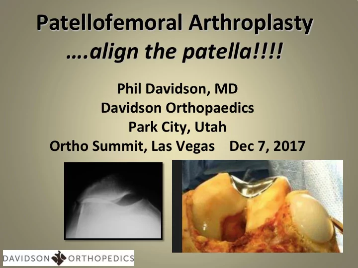
Patellofemoral Arthroplasty .align the patella!!!! Phil Davidson, MD Davidson Orthopaedics Park City, Utah Ortho Summit, Las Vegas Dec 7, 2017 Disclosures none Outline Morphology Geometry Bio vs Prosthetic


slide-1
SLIDE 1

Patellofemoral Arthroplasty ….align the patella!!!!

Phil Davidson, MD Davidson Orthopaedics Park City, Utah Ortho Summit, Las Vegas Dec 7, 2017

slide-2
SLIDE 2

Disclosures

none

slide-3
SLIDE 3

Outline

  • Morphology
  • Geometry
  • Bio vs Prosthetic
  • Prosthetics/Implants
  • Realign Procedures
  • Combo Procedures

– Realignment – Arthroplasty

slide-4
SLIDE 4

The majority of PF DJD in isolation or in “younger patients” are associated with:

  • 1. Abnormal Morphology
  • 2. Abnormal Geometry

Rotation Height Version

28 year old female

slide-5
SLIDE 5

Morphology

  • Both patellar and trochlear

morphology need to be identified in considering treatment options

  • Abnormal morphology can

create stresses on repairs

  • May not make sense to do

inlay resurfacing on abnormal morphologic topography Wiberg Classification

Dejour Classification

slide-6
SLIDE 6

Geometry

  • Geometric alignment needs

to be considered in 3-D – Patellar position M-L

  • Valgus knee

– Patellar “tilt”

  • Femoral version

– Patellar height

  • Correction targeted at

specific malalignment/rotation

  • NEVER do Lateral Release

alone

slide-7
SLIDE 7

Limb Rotation – femur and tibia

  • Both Femoral

Version AND Tibial Torsion bear on PF forces

  • Femoral Anteversion

– NL female 13

  • External Tibial

Torsion

– NL female 27

slide-8
SLIDE 8

Radiography

Merchant Xray- need dedicated board/ jig > 145 considered “shallow”

slide-9
SLIDE 9

Radiography- Patellar Height

  • Caton-Deschamps (CD)

Ratio (X/Y)

  • NL appx 0.6-1.3
  • Very handy to use

digital measuring tools

  • Patellar Alta and Baja

– Can be addressed with TTO – Alta possible MPFL

slide-10
SLIDE 10

Radiography- TT-TG or TT-PCL Relative Patellar Translation

  • TT-TG, useful if distinct

FTG sulcus

– NL <appx 18mm

  • TT-PCL, may be more

accurate, needed for abnormal FTG morphology

– NL appx <24

slide-11
SLIDE 11

Extensor Realignment

  • Medial Plication
  • Lateral Release
  • Need “normal” tissue to

plicate

– i.e. not markedly lax

  • Easily incorporated into

PFA

“Selective” lateral release, preserving underlying synovial layer– part of realignm ent, not alone!

slide-12
SLIDE 12

MPFL combo with PFA

  • This is indicated when

DJD coexists with recurrent instability and/or laxity

  • Need to protect patellar

implant

  • Avoid patellar bone

tunnel techniques

slide-13
SLIDE 13

TTO with PFA

  • Medialization can

correct for increased TT-TG or TT-PCL

  • Move proximal to

address patellar baja

  • Distalize to address

patellar alta

slide-14
SLIDE 14

Biologic or Prosthetic Resurfacing ????

Key decision making point

  • Multifactoral decision

– Lesion: focal or diffuse – Patient Factors – Comorbidities – Osteophytes – Bipolar – Resources Available

Inverted patella of 19 yr old male

slide-15
SLIDE 15

Radiographic Guide to Bio vs. Prosthetic

Kellgren-Lawrence Grading Scale

Generally Biological Resurfacing…… Grade 0 = Normal Grade 1 = Doubtful narrowing of the joint space and possible osteophytic lipping Grade 2 = Definite osteophytes, definite narrowing of the joint space Generally Prosthetic Resurfacing….. Grade 3 = Moderate multiple osteophytes, definite narrowing of joints space, some sclerosis and possible deformity of bone contour Grade 4 = large osteophytes, marked narrowing of joint space, severe sclerosis and definite deformity of bone contour.

KL 1 KL 4

slide-16
SLIDE 16

Biological Options

  • Scaffolds
  • Cell Therapy
  • Osteochondral Grafts

– Autogenous

  • Limited use

– Allograft

  • Fresh stored
  • Cryopreserved
  • Cartilage Grafts

– Minced, ground, lamellar – Cryopreserved – Non-viable (scaffold)

slide-17
SLIDE 17

Inlay vs Onlay Patella – Biologic

slide-18
SLIDE 18

What if biologics will not or cannot work? …Transitioning the algorithm from biologics to prosthetics

Prosthetics - Joint Resurfacing

slide-19
SLIDE 19

Onlay vs Inlay Joint Resurfacing - Patella

  • Inlay useful for focal

defects and for “normal” morphology

  • Onlay needed for

diffuse chondral disease or “abnormal” morphology

  • I use Onlay MUCH

more frequently

slide-20
SLIDE 20

Onlay vs Inlay Joint Resurfacing for FTG

  • Onlay device replaces

anatomy, but may add unwanted volume

  • Inlay device based on ambient

anatomy

  • Inlay device allows for

concurrent realignment

  • Inlay device inherently stable
  • Inlay typically more anatomic

Onlay

I nlay

slide-21
SLIDE 21

PFA “legacy onlay implants” very negative history

  • Non-anatomical
  • Overstuffed the joint
  • Encroach on TF joint
  • Very mixed, negative

results with high revision rates

  • Did not allow for

concurrent alignment!!

slide-22
SLIDE 22

Patellar-Trochlea alignment

Key step- Patella directly over FTG

slide-23
SLIDE 23

Summary

  • Must establish alignment
  • Want to accommodate

potential revision to TKA

  • Cannot have plastic on

cartilage (no poly patella alone)

  • Resurfacing must be done

in context of anatomy/deformities

  • No Overstuffing
slide-24
SLIDE 24

Thank You

phildavidsonmd@gmail.com