Particle Manipulation and Biosensor Applications using Optofluidic - - PDF document

particle manipulation and biosensor applications using
SMART_READER_LITE
LIVE PREVIEW

Particle Manipulation and Biosensor Applications using Optofluidic - - PDF document

Oral Presentation, Theme D : Nanobiotechnology, Nanomedicine, Nanotoxicology, Biosensors ... Particle Manipulation and Biosensor Applications using Optofluidic Ring Resonators Abdurrahman Gumus, 1,2 Sudeep Mandal 3 and David Erickson 2,4 1


slide-1
SLIDE 1

Particle Manipulation and Biosensor Applications using Optofluidic Ring Resonators

Abdurrahman Gumus,1,2 Sudeep Mandal3 and David Erickson2,4

1Department of Electrical and Computer Engineering, Cornell University, Ithaca, NY 14853, USA 2Cornell NanoScale Science & Technology Facility, Cornell University, Ithaca, NY 14853, USA 3School of Applied and Engineering Physics, Cornell University, Ithaca, NY 14853, USA 4Sibley School of Mechanical and Aerospace Engineering, Cornell University, Ithaca, NY 14853, USA

Abstract— In recent years, much advancement has been made on optofluidics field which integrates optical elements in the form of lenses, lasers, waveguides and sensors with microfluidic devices. Here, for the first time, we present the potential usage of an optofluidic ring resonators in big particle trapping and manipulation ranging in size from 10-25 μm. When light at the resonant wavelength is coupled into the bus-waveguide, optical field confined within the ring resonator becomes amplified. The resulting high optical intensities in the evanescent field of the ring enable trapping of particles on the resonator. We are also trying to investigate a novel type of biosensor that could measure the mass of a single cell by observing resonant frequency shift of the resonator. This approach could potentially provide a new optofluidic based method for mass measurements of a single adherent cell in its physiological condition in a non-invasive manner.

Many research fields benefit from the ability to manipulate particles in the micro- and nanoscale regimes. This manipulation can be achieved with a variety of forces, including mechanical, magnetic, fluidic, electrokinetic and

  • ptical forces. Optical forces which provide dynamic and

flexible manipulation can be used for micro- and nano particle manipulation either through a radiation pressure1 or by the force exerted by the gradient of the optical field of a highly focused laser beam, as in the optical tweezers2 which have been proven to be a useful tool for deflecting, sorting and transporting microparticles. Recently, researchers have developed methods such as slotted waveguides3, photonic crystal resonators4,5 and plasmonic6 structures to generate high- intensity field gradients for the trapping of nanoparticles and

  • biomolecules. Although these systems are highly advantageous

for nanoscale particle trapping, they are limited for micro scale particle trapping because nanophotonic trapping length scale is much smaller than the actual particle size. Ring resonator is a kind of planar waveguiding device that can achieve high optical intensities. It consists of a ring waveguide adjacent to a bus waveguide. Light from a laser goes through the bus waveguide and evanescently couples into the ring resonator. When the optical path length around the ring is equal to an exact multiple of the wavelength of the excitation light, ring is ON resonance. Under this condition, the intensity of the light builds up and causes a dramatic increase in the optical field confined within the ring. This highly intensive evanescent field on the resonator is being used for trapping and sensing purposes. By using tunable laser source, these resonance conditions can be detected by a corresponding drop in the power output of the bus waveguide. To be able to make resonator single mode, silicon (Si) waveguide was designed to be 450 nm wide and 250 nm tall (Fig1). Low index silicon dioxide layer (SiO2) which lies beneath the high index Si waveguide helps to confine the light within the waveguide core. Silicon-on-insulator wafers were patterned using electron-beam (e-beam) lithography and etched using an inductively coupled plasma etching system. 1.8 μm of SiO2 was evaporated onto the chip using e-beam evaporation. Lift off processing was used to mask each resonator on the chip while SiO2 evaporation. Each ring resonator has different radius and spacing to the bus waveguide. Since the resonant wavelength is dependent on these properties, each resonator has a unique resonant wavelength associated with it. In summary, we have demonstrated an optofluidic ring resonator device that can be used for micro scale particle trapping and manipulation. We present the potential usage of these devices for trapping of big planar particles ranging in size from 10-25 μm. We are also investigating a novel type of

  • ptofluidic biosensor that could measure the mass of a single

cell by observing resonant frequency shift of the resonator. We are also planning to work on developing label-free biosensing applications by combining this optically driven ring resonator device with polymer brushes.

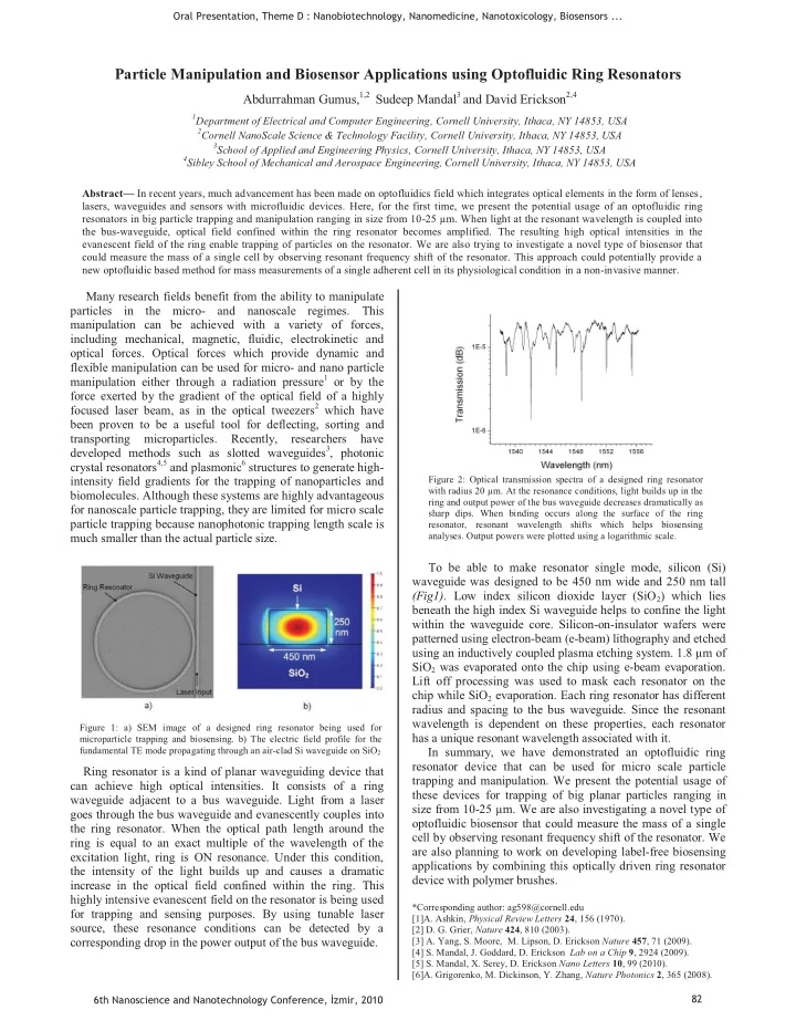
*Corresponding author: ag598@cornell.edu [1]A. Ashkin, Physical Review Letters 24, 156 (1970). [2] D. G. Grier, Nature 424, 810 (2003). [3] A. Yang, S. Moore, M. Lipson, D. Erickson Nature 457, 71 (2009). [4] S. Mandal, J. Goddard, D. Erickson Lab on a Chip 9, 2924 (2009). [5] S. Mandal, X. Serey, D. Erickson Nano Letters 10, 99 (2010). [6]A. Grigorenko, M. Dickinson, Y. Zhang, Nature Photonics 2, 365 (2008). Figure 2: Optical transmission spectra of a designed ring resonator with radius 20 μm. At the resonance conditions, light builds up in the ring and output power of the bus waveguide decreases dramatically as sharp dips. When binding occurs along the surface of the ring resonator, resonant wavelength shifts which helps biosensing

  • analyses. Output powers were plotted using a logarithmic scale.

Figure 1: a) SEM image of a designed ring resonator being used for microparticle trapping and biosensing. b) The electric field profile for the fundamental TE mode propagating through an air-clad Si waveguide on SiO2

Oral Presentation, Theme D : Nanobiotechnology, Nanomedicine, Nanotoxicology, Biosensors ... 6th Nanoscience and Nanotechnology Conference, zmir, 2010 82

slide-2
SLIDE 2

Bioactivation of Titanium Based Orthopedic Implants via Titania Nanotubes

Cem Bayram,1,2 Murat Demirbilek1, Nazlı Çalışkan1, Melike Erol Demirbilek3, Tayfun Vural4 and Emir Baki Denkbaş*1

1Nanotechnology and Nanomedicine Division, Hacettepe University, Ankara 06800, Turkey

2Department of Chemistry, Aksaray University, Aksaray 68100, Turkey 3 Department of Biology, Aksaray University, Aksaray 68100, Turkey 4Department of Chemistry, Hacettepe University, Ankara 06800, Turkey

Abstract— In this study, we present that medical grade titanium based plaques were oxidized with anodic oxidation in electrolyte solution. The resulting oxide layer is tube patterned with approximately 50 nm in diameter. Anodized titanium plaques were immersed into vancomycin containing simulated body fluid in order to load with antibiotics. The cell culture and related in vitro tests were carried out. Titania nanotube formation affected positively in osteoblast proliferation and bacterial inhibition.

Orthopedic implants have been largely used in people in both developed and developing countries with the prolonged lifetime. Complications such as bacterial infections and insufficient

  • sseointegration

are the primary disadvantages of post implantation. If not prevented, bacterial infection can result in serious and life threatening conditions such as osteomyelitis [1]. Although titanium and titanium alloys are all cytocompatible and show proper mechanical strength, in order to reduce chances of such serious complication, patients are often subjected to antibiotic drug therapy for 6–8 weeks after initial surgery. The antibiotics are systemically delivered either intravenously, intramuscularly or topically. Systemic antibiotic delivery entails certain drawbacks such as systemic toxicity. The local presence of antibiotics prevents the body from the disadvantages mentioned above [2]. If we think bone as a nanocomposite which consists of

  • rganic and inorganic constituents (such as collagen

nanofibers and nano sized hydroxyapatite crystals) we can conclude that the best integration can be achieved with nano structured biomaterial surfaces. The titania nanotube pattern showed better integration in previous studies [3]. The titanium alloy plaque samples were oxidized with size controlled titania layer by anodization method. In a typical procedure titanium plaques with the diameters of 10 mm are used as anode and platinium mesh structure was used as cathode. The electric potential was kept constant and the procedure lasted for 10 min. Vancomycin hydrochloride was chosen as antibacterial agent and was incorporated into apatite crystals The plaques were then immersed in vancomycin hydrochloride containing 1.5X simulated body fluid (SBF) solution for 3 days [4]. The characterization of anodization and apatite formation procedures were carried out by AFM and SEM techniques. The release of drug from apatite crystals was determined spectrophotometrically.

Figure 1 and 2. Anodization setup and SEM image of anodized Ti6Al4V plaque.

Human osteosarcoma Saos-2 /An1 cell lines was used cell culture experiments. Cell cultures were conducted in sterile 24-well TCPS dishes in stationary conditions. Modified and antibiotic loaded titanium implants, having 10 mm diameter and 5μm thickness, were placed in 24-well TCPS dishes, fifty microliters of cell suspension (2x10³ cell/well) was pipeted onto the each implant then incubated in a humidified incubator. Adhesion and proliferation of Saos- 2/An1 on the titanium implants were analyzed by crystal violet staining assay, after different culture times 2,5, 7 and 10 days. The absorbance at 570 nm was measured. Osteoblast adhesion and proliferation studies results showed that titania nanotube modification did increase

  • steoblast

attachment and proliferation (p<0,05). The amount of total protein levels were measured 3,5,7 and 10 days of cultured cells onto the modified and non- modified titanium implants by Lowry’s method. Alkaline phosphatase activity (ALP) was measured to the cells cultured onto the implants for 5,7 and 10 days. The cells present modified implant surfaces showed higher ALP levels compare to on the non modified titanium surfaces [5]. (p<0.05). To evaluate the effect of surface modification and vancomycin on the matrix mineralization of Saos-2/An1, Alizarin Red staining of cell cultured on the implants was carried out, than photographed by the light microscopy. Growth of microorganisms in direct contact with the biomaterial surface plays a pivotal role in biofilm formation. Thus, in this work we have investigated bacterial adhesion on nanotubes loaded with vancomycin. A bacterial cell line Staphylococccus aureus ATCC 25923 was used in the experiments. Data are expressed as means ± standard deviations of a representative of three similar experiments carried out in

  • triplicate. Statistical analysis was performed using the

Statistical Package for the Social Sciences (SPSS) version 11.5 software. Statistical comparisons were made by analysis

  • f variance (ANOVA). In all statistical evaluations, p<0.05

was considered as statistically significant.

*Corresponding author: denkbas@hacettepe.edu.tr [1] Nandy et al., Mat Sci Eng C 29 2478 (2009). [2] Jose et al., Chem & Bio, 12 1041 (2005) [3] Oh et al.,J Biomed Mater Res 78A 97 (2006) [4] Yao and Webster, JBiomed Mater Res Part B :Appl Biomater 91B 587 (2009) [5] Popat et al., Biomaterials 28 4880 (2007) Oral Presentation, Theme D : Nanobiotechnology, Nanomedicine, Nanotoxicology, Biosensors ... 6th Nanoscience and Nanotechnology Conference, zmir, 2010 83

slide-3
SLIDE 3

Electrochemical Detection of Influenza A Virus based on DNA Hybridization by using Voltammetric and Impedance Biosensors

Dilsat Ozkan-Ariksoysal

1*, Buket Meric1, Nilay Aladag1, Mehmet Ozsoz1

1Ege University, Faculty of Pharmacy, Analytical Chemistry Department, 35100, Izmir, Turkey

Abstract-The aim of this study was to develop a novel electrochemical genosensor for the detection of DNA sequences related to the Influenza A virus genotype based on the intrinsic guanine oxidation signal. Easy wet-adsorption method was used to provide immobilisation of label-free probe DNA related to the Influenza A virus onto disposable carbon graphite electrode (GCE) surface without using any additional chemical agent for the immobilisation. The probe-modified sensor was able to clearly distinguish perfect match from non-complementary DNA in only 15 min. detection time. The voltammetric data were also confirmed and compared with electrochemical impedance spectroscopy (EIS). Several factors affecting on the hybridization and the rate of fullmatch/non- complementary discrimination are studied to maximize sensitivity and selectivity.

It has been known that a significant public problem of Influenza viruses cause three to five million severe cases in each year and between 250,000 and 800,000 deaths according to WHO data [1] These viruses may bring about such important diseases such as pneumonia, which can be fatal both adults and children and they can also cause economic loss[2]. The rate of influenza infections and excess deaths increased in each year. For all these important reasons, detection of the Influenza viruses is of importance in the medical and social area. Several techniques have been reported for the detection of Influenza as viral culture, immunofluorescence staining (IFA), enzyme immunoassays (EIA), RT-PCR, DNA microarrays, and Taq man based real time PCR. The classical viral culture method is slow and it takes from 2 to 14 days and others have such disadvantages that immunofluorescence staining technique requires intact cells, highly skilled research man and specialised equipment, enzyme immunoassay is high cost and it is not a virus recoverable method. Although, PCR method have stil been used for the sensitive and molecular detection of many viruses, after the amplification of selected gene region, toxic chemical as ethidium bromide is used in agarose gel electrophoresis step. There are also some rapid tests used for the detection of influenza viruses and the rapid detection of Influenza viruses are important for taking preventative measures, changing the anti-viral theraphy. However, the reliable discrimination of the influenza virus type is not possible with them because these tests either detect and distinguish between influenza A and B or detect influenza A only. The alternative biological material detectors “biosensors” have been developed during the past two decade to fill a need for simple, rapid, inexpensive and portable testing devices for identifying biomolecular interactions. In these field, many DNA biosensors have been reported for the detection of DNA hybridization, DNA mutations, drug- DNA interactions by using optical, piezoelectric and electrochemical transducers recently [3,4]. The electrochemical detection

  • f

speci

  • hybridization events[5,6] can be monitored based on the

intrinsic guanine signal or by mostly DNA hybridization indicators as antibiotics or some metal complexes [7-9] or by using some nano-sized materials[10]. Here, we described a simple, label-free and inexpensive voltammetric and impedimetric DNA biosensors for the detection of Influenza A viruses as an alternative to traditional methods and biosensors which were early described in the literatures. The detection of the hybridization event was evaluated based on guanine

  • xidation signal for voltammetric biosensor and it was also

monitored in the presence of [Fe(CN)6]3-/4- based on electron transfer resistance (Rct). After addition of the complementary oligonucleotide onto the probe-modified GCE, responses obtained from both sensors showed maximum increase. The specificity and detection limit of biosensors were detected differences/ advantages of both protocols were discussed. Additionally, preliminary results

  • f

electrochemical detection and discrimination

  • f

Influenza A and B viruses from PCR samples based on the guanine signal were also evaluated. (a) (b)

Figure 1. Differential pulse voltamogram (a) and impedance spectra (b) of the DNA hybridization related to Influenza A virus.

The work was supported by TUBITAK (Project No: SBAG-105S484). *Corresponding author: dilsat.ariksoysal@ege.edu.tr

[1]http://www.who.int/mediacentre/factsheets/fs211/ en/index.html. [2]Ward C.L., M.H. Dempsey, C.J.A. Ring , R.E. Kempsond, Journal of Clinical Virology 29, 179, (2004). [3] Palecek E.. Past, presence and future of nucleic acids

  • electrochemistry. Talanta, 56: 809-82, (2002).

[4] Wang, J. Anal. Chim. Acta, 2002, 469, 63-71. [5] Ozkan-Ariksoysal, B. Tezcanli, B. Kosova, M. Ozsoz, Anal. Chem., 80, 588-596 (2008). [6] Long, Y.T.; Li, C. Z.; Sutherland, T.C.; Kraatz, H.B.; Lee, J.

  • S. Anal. Chem., 76, 4059-4065, (2004).

[7] Erdem, A., Kerman, K Meric, B., Akarca, U.S., Ozsoz, M.

  • Anal. Chim. Acta, 422, 139–149, (2000).

[8] Kelley, S. O.; Boon, E.M.; Barton, J. K.; Jackson, N. M.; Hill,

  • M. G. Nucl. Acids Res., 27, 4830-4837, (1999).

[9] Ozkan, D., Erdem, A., Kara, P., Kerman, K., Gooding, J. J., Nielsen, P. E., Ozsoz, M. Electrochem. Commun., 4 (10), 796- 802, (2002). [10] Ozsoz M, Erdem A., Kerman K., Ozkan D., Tugrul B., Topcuoglu N., Ekren H., Taylan, Anal.Chem., 75, 2181-2187, (2003).

Oral Presentation, Theme D : Nanobiotechnology, Nanomedicine, Nanotoxicology, Biosensors ... 6th Nanoscience and Nanotechnology Conference, zmir, 2010 84

slide-4
SLIDE 4

Emerging Nanotechnology tools for drug delivery: Carbon nanotubes

İ. Tuncer DEĞİM, Sibel İLBASMIŞ - TAMER

Gazi University, Faculty of Pharmacy, 06330 Etiler, Ankara, TURKEY Abstract- Nanotechnology is rapidly developing field in especially engineering and medical sciences. Carbon nanotubes are one of the most studied nanomaterials in material sciences and physics but not in medical sciences and pharmacy. The adsorption and desorption properties of carbon nanotubes were investigated for ibuprofen, naproxen, oxaliplatin and paclitaxel. Multiwalled carbon nanotubes were also PEGylated and teh effect of PEGylation was investigated.

Carbon nanotubes are first proposed to the market in 19911 and appeared to be a new and emerging nanotechnology tool for drug delivery in nanomedicine. Some of nanotechnology applications can be tailored early diagnosis and treatment of many diseases including some skin disorders and may be

  • cancers. If nanoparticles are able to penetrate through skin

layers or if they can be able to provide drug molecules on to the skin surface they can be used to deliver active substances for therapeutic purposes but exploration

  • f

their pharmaceutical applications remains at a very early stage2. Recently it has been noticed that single-walled carbon nanotubes (SWNTs) and multi walled carbon nanotubes (MWNTs) can be internalized by living cells and can pass across the biological membranes in cell culture studies3. The internalization of carbon nanotubes by corneocytes has been shown4 in the literature. Figure 1 shows subsequent desorption of NAP and IBU after adsorption. Figure 1. Desorption of NAP and IBU from CNTs after adsorption (24h). Figure 2. Penetrated amount of drugs through full thickness rat skin. Skin penetration experiments using CNTs and drug complexes were also done and higher penetrations were

  • bserved (Figure 2). Higher penetrations were observed when

drugs were provided with CNTs. The physical appereance of MWCNTs were determined using AFM (Figure 3). The tubular shapes of CNTs were clearly

  • seen. On the figure and branches of CNTs and carbon

particles can be observed. This gives information mainly about the lenth of nanotubes and approximate valuation of the bundles diameter. It was found not to be very accurate to determine exact value

  • f diameter of CNTs as it was reported in the literature5

because some nanotube does not lie directly on a mica and

  • scilated under the daze.

Figure 2. AFM images of DWCNTs and MWCNTs Figure 3. AFM images of PEGylated MWCNTs. Figure 3 shows the effect of PEGylation. The diameter was found to be increased after PEGylation. As a conclution to prepare CNTs-drug mixture is possible and it is resulted in drug adsorption. CNTs can adsorp drug molecule on their surface and it can deliver the drug and even they can release the drug by subsequent desorption. CNTs were found to be suitable material for drug transport having higher surface area and being an adsorptive material. PEGylation process was found to be effective and useful technique for further studies. *tunc@tr.net

1-S. Iijima, Nature 354:56 (1991). 2- C.R. Martin, P. Kohli, Nat. Rev. Drug Discov. 2:29 (2003). 3-L. Lacerdaa et al., Nanotoday 2:38 (2007). 4-A. N. Monterio-Riviere et al., Nanotoxicology 1:293 (2005). 5- K. Safarova et al., Modern Research and Educational Topics in

  • Microscopy. 513-519 (2007).

Oral Presentation, Theme D : Nanobiotechnology, Nanomedicine, Nanotoxicology, Biosensors ... 6th Nanoscience and Nanotechnology Conference, zmir, 2010 85

slide-5
SLIDE 5

Interferometric Reflectance Imaging: Nanoscale Biological Imaging, Label-free Protein and Single Pathogen Detection

  • M. Selim Ünlü

Electrical and Computer Engineering, Center for Nanoscience and Nanobiotechnology, Boston University, Boston, MA 02215, USA Abstract — Utilizing the sensitivity provided by optical interference, we have demonstrated nanometer scale measurement capability in fluorescence microscopy studying conformation of DNA protein complexes. We have also developed a novel platform based on Interferometric Reflectance Imaging Sensing (IRIS) capable of high-throughput label-free biodetection in a micro-array format. The IRIS technique relies on a layered surface and imaging the reflectance when the sensor surface is illuminated, for example by a tunable laser, at different wavelengths. We have demonstrated dynamic multiplexed detection of antigens and antibodies in solution using corresponding probes on the IRIS surface as well as label-free measurements of DNA hybridization kinetics and single pathogen detection capability. Optical interference is one of the key properties of light enabling important devices such as interference filters and lasers. We have utilized basic principles of optical interference in biological applications demonstrating nanometer scale measurement capability in fluorescence microscopy and label-free sensing of protein binding in a high-throughput micro-array format. A novel application of interference to fluorescence microscopy promises nanometer resolution in biological imaging. We have developed a technique – spectral self-interference fluorescent microscopy (SSFM) – that transforms the variation in the intensity for different wavelengths in emission of fluorophores on a layered surface to nanoscale height information. Using SSFM, we have estimated the shape of coiled single-stranded DNA, the average tilt of double-stranded DNA of different lengths, and the amount of hybridization [1]. The determination of DNA conformations on surfaces and hybridization behavior provide information required to move DNA interfacial applications forward and thus impact emerging clinical and biotechnological

  • fields. Recently, we have also applied SSFM to study the

conformational changes of polymers [2] and DNA-protein

  • complexes. [3] We combined SSFM with a complementary

technique invented by Prof. Rant at Technical University of Munich (TUM) in which surface electric field provides highly-

  • rdered DNA arrays [5] and SSFM can quantify conformational

changes when DNA-protein complexes form. Direct monitoring of primary molecular binding interactions without the need for secondary reactants would markedly simplify and expand applications of high-throughput label-free detection methods. We developed Interferometric Reflectance Imaging Sensing (IRIS) that monitors the optical phase difference resulting from accumulated biomolecular mass and demonstrated simultaneous detection of antigens and antibodies. [6] Dynamic measurements were made at ~10 pg/mm2 sensitivity. We have also demonstrated label-free measurements of DNA hybridization

  • kinetics. [6] Recently, we have developed a multi-LED discrete

wavelength illumination system that allows for high spatial resolution imaging in IRIS modality. We have used this system to detect nanoscale particles and characterize their reflection signature thus measuring the size of the particle [7]. We have successfully detected 100 nm and 70 nm particles.

Funding for this research was provided by NIH/NIGMS (R21 GM074872-02), US Army (W911NF-06-0040), NSF (CBET-0933670 and OISE-0601631) and Smart Lighting Engineering Research Center (EEC-0812056). *Corresponding author: selim@bu.edu [1] L. Moiseev, et al., “DNA Conformation on Surfaces Measured by Fluorescence Self-Interference,” PNAS, 103, pp. 2623–2628, (2006) [2] A. Yalcin, et al.,, “Direct Observation of Conformation of a Polymeric Coating with Implications in Microarray Applications,” Anal. Chem., DOI: 10.1021/ac801954x (2009) [3] P. S. Spuhler, et al., “Platform for in situ real-time measurement of protein-induced conformational changes of DNA,” PNAS, January 2010 [4] U. Rant et al., “Dynamic electrical switching of DNA layers on a metal surface,” Nano Letters, 4:2441-2445, (2004) [5] E. Ozkumur, et al., “Label-free and dynamic detection of biomolecular interactions for high-throughput microarray applications,” PNAS, 105, pp. 7988–7992 (2008). [6] E. Ozkumur, et al., “Label-free microarray imaging for direct detection of DNA hybridization and single-nucleotide mismatches,” Biosensors and Bioelectronics, Vol. 25, No. 7 (2010). [7] G. G. Daaboul, et al., “LED-based spectral reflectance imaging biosensor for label-free high-throughput multi-analyte and single-pathogen detection,” SPIE Photonics West 2010 - BIOS, January/February 2010

  • Fig. 1: (left) Rant group orients the DNA on a gold surface by applying

electrostatic potential. Adding a transparent metal film on the top surface

  • f the layered structure for SSFM (right) allows for orienting the ds-DNA

as a uniform brush and thus any conformational changes when DNA forms a complex with a protein can be directly measured.

h h’

Silicon Substrate

Layered structure ITO h h’ h h’

Silicon Substrate

Layered structure ITO

  • Fig. 2. Clockwise from the bottom (a) IRIS platform consists of an oxide

surface on a Si substrate. Biomolecule accumulation on the surface changes the optical path difference between the top surface and buried

  • xide-Si interface. (b) Reflection spectroscopy with a tunable illumination

interrogates layer thickness. The sample reflectance is recorded as an intensity image by a camera (c) forming a reflectivity-vs.-wavelength curve at each pixel of the CCD. The thickness at each pixel (thus binding) is found by processing these recorded curves. (d) Prototype instrument. Oral Presentation, Theme D : Nanobiotechnology, Nanomedicine, Nanotoxicology, Biosensors ... 6th Nanoscience and Nanotechnology Conference, zmir, 2010 86

slide-6
SLIDE 6
  • Oral Presentation, Theme D : Nanobiotechnology, Nanomedicine, Nanotoxicology, Biosensors ...

6th Nanoscience and Nanotechnology Conference, zmir, 2010 87

slide-7
SLIDE 7

Electrochemical monitoring of DNA hybridization by magnetic particles based sensor technology

Hakan Karadeniz1, Arzum Erdem1*

1Ege University, Faculty of Pharmacy, Analytical Chemistry Department, Izmir 35100, Turkey

Abstract-An indicator-based and indicator-free magnetic assays connected with a disposable pencil graphite electrode (PGE) were successfully developed, and also compared for the electrochemical detection of DNA hybridization. The oxidation signals of echinomycin (ECHI) and electroactive DNA bases, guanine and adenine, respectively were monitored in the presence of DNA hybridization by using differential pulse voltammetry (DPV) technique. The detection limits (S/N = 3) of the magnetic assays based on indicator or indicator-free were found in nM concentration level of target using disposable sensor technology with good reproducibility.

The development of advanced biological sensor systems could impact signicantly the areas of genomics, proteomics, biomedical diagnostics and drug discovery [1,2]. Recent advances in biosensors based on nucleic acid have led to the development of genosensor technology for gene sequence analysis and for nucleic acid ligand binding studies [3-5]. The electrochemical nucleic acid sensor system based on magnetic particles [6-11], labeling with an enzyme [8], using label-free system [7,9,11] or combining with metal nanoparticles [6,10], enables the sequence specic detection of DNA hybridization observed in low detection limits resulting from ef

  • A biomagnetic assay of DNA sequences related to breast-

cancer gene (BRCA1) was reported based on label-free detection [7]. Coupling the label-free guanine detection with the ef biotinylated inosine-substituted probes, streptavidin-coated magnetic beads with potentiometric stripping analysis (PSA) measurements at a renewable graphite pencil electrode. In this study, an indicator-based and indicator-free magnetic assays connected with a disposable graphite sensor (pencil graphite electrode, PGE) were successfully developed, and also compared for the electrochemical detection of DNA

  • hybridization. The changes at the oxidation signals of

echinomycin (ECHI) and electroactive DNA bases, guanine and adenine, respectively were monitored in the presence of DNA hybridization by using differential pulse voltam- metry (DPV) technique. The selectivity of these magnetic assays for DNA hybridization was also checked in the presence of single base mismatch and noncomplementary DNA sequences. There have not been yet any reports about both indicator-based and indicator-free magnetic assays connected with disposable sensor system by measuring the oxidation signals of ECHI, guanine and adenine in the same measurement scale. The reported magnetic assays based on indicator-based and indicator-free methods connected with a disposable graphite sensor (PGE) were successfully developed for the electrochemical detection of DNA hybridization. There have not yet been any reports of both indicator-based and indicator-free magnetic assays connected with a disposable sensor system through measurement of the oxidation signal of indicator ECHI with the signals of DNA bases, guanine and adenine, in the same measurement scale. Easier, quicker and

more

sensitive detection sche me for DNA

hybridization based on magnetic assay was e xplored herein in co mparison to the traditional techniques reported earlier in

the literature [5,12], in which several e xternal indicators [Co(phen)3]3+, methylene blue, meldola’s blue have been applied by using advanced surface modi

  • r

regeneration

sche mes. The use of PGE brings the other advantage to our assay with a better reproducibility; such as, being cheaper, easy to use (single-use) and portable, which are crucial properties of devices for DNA chip technology contrary to other transducers; such as, gold electrode and hanging mercury drop electrode. A.E. acknowledges the financal support from TUBITAK (Project no.106S181). H.K. acknowledge a scholarship from PhD students obtained from TUBITAK. *Corresponding author: arzum.erdem@ege.edu.tr

[1] P.M. Dean, E.D. Zanders, D.S. Bailey, Trends Biotechnol. 19 (2001) 288. [2] J. Wang, Analyst 130 (2005) 421. [3] E. Palecek, F. Jelen, F. Crit. Rev. Anal. Chem. 32 (2002) 261. [4] A. Erdem, M. Ozsoz, Electroanalysis 14 (2002) 965. [5] A. Erdem, K. Kerman, B. Meric, U.S. Akarca, M. Ozsoz,

  • Anal. Chim. Acta 422 (2000) 139.

[6] J. Wang, D.K. Xu, R. Polsky, J. Am. Chem. Soc. 124 (2002) 4208. [7] J. Wang, A.N. Kawde, A. Erdem, M. Salazar, Analyst 126 (2001) 2020. [8] J. Wang, D.K. Xu, A. Erdem, R. Polsky, M.A. Salazar, Talanta 56 (2002) 931. [9] A. Erdem, D.O. Ariksoysal, H. Karadeniz, P. Kara, A. Sengonul, A. Sayiner, M.Ozsoz, Electrochem. Commun. 7 (2005) 815. [10] J. Wang, G.D. Liu, R. Polsky, A. Merkoci, Electrochem.

  • Commun. 4 (2002) 722.

[11] H. Karadeniz, A. Erdem, F. Kuralay, F. Jelen, Talanta, 78 (2009) 187. [12] A. Erdem, K. Kerman, B. Meric, U.S. Akarca, M. Ozsoz, Electroanalysis 11 (1999) 586.

Oral Presentation, Theme D : Nanobiotechnology, Nanomedicine, Nanotoxicology, Biosensors ... 6th Nanoscience and Nanotechnology Conference, zmir, 2010 88

slide-8
SLIDE 8

Direct Electrical Detection of E.Coli Based on Different DNA Modified Genosensors

i,1, 1, P. Kara1*, A. Yurtman2 and M. Özsöz1

1Department of Analytical Chemistry, Faculty of Pharmacy, Ege University, 35100, Bornova, Izmir, Turkey 2

Department ofMicrobiology, Faculty of Pharmacy, Ege University, 35100, Bornova, Izmir, Turkey Abstract— Herein, we describe an impedimetric and voltammetric genosensor for rapid and direct electrochemical detection of E.Coli PCR (Polymerase Chain Reaction) products by using different modification techniques. Graphite and gold sensor surfaces were modified with probe sequences via different modification techniques. The aim of the study is to develop nanosensors for direct and rapid detection of microorganisms.

Recent advances in biosensors based on nucleic acid hybridization recognition have led to the development of genosensor technology for DNA sequence analysis.[1-3] Specifically, electrochemical hybridization biosensors demonstrate great promise for pathogen identification, mutation detection, and genomic sequencing[4-5]. These novel, sequence-specific hybridization processes either involve monitoring the oxidation signal of the electroactive bases of DNA [6-7]

  • r employing an electroactive

hybridization indicator [8-9] that emits different signals to discriminate between single-stranded and double-strand DNA. Successful attempts to exploit the electrochemical detection of hybridization events and base pair mismatches using sample amplicons in order to obtain reliable, clinically-relevant measurements have already been reported. Genosensors clarify the array technologies which based on immobilized 15–25 mer single strand DNA called as capture probe molecules [10]. These probe molecules are the complementary sequences of the specific part of the amplicon related to a virus, bacteria or a specific gene sequences. The specificity and the selectivity of the hybridization detection by using long amplicons are dependent on the conditions at the electrode surface such as; density, extent, and relative position

  • f probe sequence.

In this work effect of surface chemistry onto hybridization and detection of microbiological disease was performed. E. coli probe and PCR products were used as a model case. For this purpose, disposable graphite and gold surfaces were modified by using a variety of different methods such as adsorption, covalent agent, poly-D-lysine, trimethoxysilane, carbon nanotubes (CNT), gold nanoparticles (AuNP) and streptavidene/biotin. The aim of this study is to select the most appropriate surface modified electrode for hybridization and clinical detections of microorganisms. Inosine substitute probe sequence representing E.Coli bacteria was immobilized on the modified electrodes and the hybridization process

  • ccurred.

Namely, DNA probe underwent hybridization to its target, mismatch and non- complementary oligonucleotides. Hyridiziation and mismatch detection was performed by electrochemical impedance spectroscopy and g differential pulse voltammetric signals of guanine. There was also an enhancement in the hybridization signals due to the increased amount of immobilized oligonucleotides

  • n the electrodes when it was compared to hybridization

signal of unmodified electrodes *Corresponding author:

[1] H.H. Thorp, Trends in Biotechnology 16, 117, (1998). [2] S. Sawata, E. Kai, K. Ikebukoro, T. Iida, T. Honda, I. Karube, Biosensors&Bioelectronics 14, 397, (1999). [3] M. Ozsoz, A. Erdem, D. Ozkan, P. Kara, H. Karadeniz, B. Meric,

  • K. Kerman, Bioelectrochemistry 67, 199, (2005).

[4] D. Ozkan, A.Erdem, P. Kara, K. Kerman, B. Meric, J. Hassmann,

  • M. Ozsoz,

[5]F. Lucarelli, G. Marazza, I. Palchetti, S. Cesaretti, M. Mascini,

  • Anal. Chem. 74, 5931, (2002).

[6] . Wang, A.N. Kawde,

  • Anal. Chim. Acta 469, 93, (2002).

[7] P. Kara, C. Cavusoglu, S. Cavdar, M. Ozsoz, Biosensors& Bioelectronics, 24, 1796, (2009).

  • Anal. Chim. Acta 431, 219, (2001).

[8] E [9] . Palecek, M. Fojta, M. Tomschik, J. Wang, Biosensors&Bioelectronics 13, 621, (1998). [10] P. Kara, S. Cavdar, A. Berdeli, M. Ozsoz, Electroanalysis, 19, 1875, (2007).

  • P. Kara, B. Meric, A. Zeytinoglu, M. Ozsoz, Anal. Chim. Acta

518, 69, (2005).

Oral Presentation, Theme D : Nanobiotechnology, Nanomedicine, Nanotoxicology, Biosensors ... 6th Nanoscience and Nanotechnology Conference, zmir, 2010 89

slide-9
SLIDE 9

Detection of Mycobacterium tuberculosis complex and Mycobacterium gordonae on the same portable surface plas mon resonance sensor

Memed Duman1* and Erhan Piskin1

1Hacettepe University, Chemical Engineering Department and Bioengineering Division, and R&D Center for Bioengineering and Biyomedtek,

Ankara, 06800 Turkey Abstract-In the present study, we have developed specific detection system for M. tuberculosis complex and M. gordonae by using a commercially available SPR based portable-multichannel sensor system. The results showed that sensor platform can be used to detect DNA hybridization effectively down to a concentration of 0.05 . The sensor platform can be regenerated with 2.5 mM HCl quite effectively and reused several times without losing the signal intensity. The SPR sensors carrying the probe-ssODNs were kept in vacuum at room temperature in the dark for about 12 weeks, and used effectively.

Tuberculosis (TB) is a common and deadly infectious disease caused by several mycobacteria especially Mycobacterium tuberculosis complex (MTB) consisting of M. tuberculosis, M. bovis, M. africanum, M. microti. According to World Health Organization, [1] globally, there were about 1.7 million deaths from TB in 2007 occurring world-wide. On the other hand, Mycobacterium gordonae, typically regarded as a colonizing organizm, is a nontuberculous mycobacterium (NTM) which are found primarily in natural and tap water, but are also found in soil, dust, animals, and food. Moreover, the presence of M. gordonae in hospital water system and other environments causes contamination and detected as a false positive result in many conventional TB test [2]. Therefore, distinguish of M. gordonae among MTB complex species is essential. Surface Plasmon Resonance (SPR) is a rather new optical technique which allows following the interactions between biomolecules without using any label. It has been applied to the measurements of ligand-receptor interactions, drug screening, DNA-hybridization [3], enzyme-substrate interactions, polyclonal antibody characterization, epitope mapping and label free immunoassays. In the present work, we developed a three channel mini (portable) SPR system for label free detection of M. tuberculosis complex and M. gordonae. A thiolderivatized single-strand oligodeoxynucleotides (ssODN), which are complementary of the target characteristic sequence of MTB complex and M. gordonae were used as the “probes” and immobilized different channel of SPR chip. A probe- immobilization protocol was developed and the sensor carrying the probes as nano-overlayers was used for the detection of the both “target” ssODNs and PCR products of M. tuberculosis complex and M. gordonae. A non complementary ssODNs was also used as control to exhibit the selectivity of the sensor. The prepared sensor performances and calibration curves were examined for detection of both M. tuberculosis complex and M. gordonae and a steady increase in the SPR signal was

  • bserved when the target ssODNs concentration increased up

1 M and the maximum relative refractive index was measured 82 ± 8.2 × 10 RIU and 146 ± 13 × 10 Cross-selectivity of the SPR sensors was tested to determine the MTB complex in mixed sample solutions that contained both MTB target and non-complementary sequences. Figure 1 presents changes in the refractive index responses in all 3 channels of SPR sensor chips when they were incubated with mixed sample solution. As expected, we just observed increase in relative refractive index in channel A which was carrying only the MTB-probe-ODNs. RIU,

  • respectively. Note that the change of the relative refractive

indices with the M. tuberculosis complex target concentration was linear only up to 0.5 M with a linear regression coefficient of 0.962. A calibration curve of M. gordonae was also performed when the target ODNs concentration in 0-2 M and linear relationship can be observed up to 1 M with a regression coefficient of 0.989.

2 4 6 8 10 12 14 16 18 20 20 40 60 80 100

Ti ( i t ) Channel A Channel B Channel C

Channel A Channel B Channel C 2 4 6 8 10 12 14 16 18 20 20 40 60 80 100

Ti ( i t ) Channel A Channel B Channel C

Channel A Channel B Channel C

Figure 1. Changes in the refractive index responses in channel A (blue triangle, ), B (red square, ) and C (black circle,

  • ) which

were modified with MTB-probe-ssODNs, GOR-probe-ssODNs and

  • nly MHC molecules, respectively.

The results showed that sensor platform can be used to detect DNA hybridization effectively down to a concentration

  • f 0.05 M and can also be used quite specifically in aqueous

solutions at room temperature and by circulation the test solution for about 20 min at a flow rate of 5 l min *Corresponding author: memi@hacettepe.edu.tr to effect complete hybridization. It was also demonstrated that the sensor platform can be regenerated with 2.5 mM HCl quite effectively and reused several times without losing the signal

  • intensity. The shelf-life can be considered acceptable when the

platforms carrying the probe-ssODNs are kept in vacuum at room temperature in the dark for about 12 weeks.

[1] World Health Organization, Global Tuberculosis control 2009 report. [2] P.B. Eckburg et al, Chest 117:96–102 (2000). [3] M Duman et al, Sensor Letters 7(4):535-542 (2009).

Relative refractive index (RIU x 10-6) Time (minutes)

Oral Presentation, Theme D : Nanobiotechnology, Nanomedicine, Nanotoxicology, Biosensors ... 6th Nanoscience and Nanotechnology Conference, zmir, 2010 90

slide-10
SLIDE 10

A B C D E Figure 1: (A) Polymeric nanoparticles loaded with bioactive agent and conjugated with biological molecules, (B) Liposomes, (C) Dendrimers, (D) Carbon nanotubes, (E) Modified surfaces with bioactive molecules A B C Figure 2: Polyurethane surfaces (A) Control, (B) Heparin modified, (C) After treatment with blood Figure 3: SEM images; (A) HAp loaded scaffold, (B) SaOs cells on the surface

Nano Alterations of Macro Systems in Biomedicine

Nesrin Hasirci1,2,3,4,5

Middle East Technical University, Faculty of Arts & Sciences, 1Department of Chemistry, Graduate Departments of 2Biomedical Engineering, 3Biotechnology, 4Micro and Nanotechnology, 5Polymer Science and Technology, Ankara 06531, Turkey nhasirci@metu.edu.tr Abstract— Polymers are commonly used in medial area in the production of implants and prostheses where the biodegradable and biocompatible natural originated polymers are the desired ones for the preparation of drug carrier devices and tissue engineering scaffolds. Micro and nano modifications of the bulk or the surface of the prepared systems may be needed to enhance their biocompatibility. This presentation will focus on the types of nano modifications of the medical systems.

Polymers are the most preferable materials in medical applications because of their versatility and ease of synthesis. They are used in the production of blood bags, artificial veins, dental fillings, contact lenses, coatings for metallic implants,

  • etc. Biodegradable and biocompatible natural originated

polymers are the ones used for the preparation of scaffolds for tissue engineering, drug carriers as micro and nano capsules, hydrogels as filling materials. Scaffolds with fibrous or porous structures have walls with different chemistry, physical shape, crystallinity, topography and surface free energy, and all these factors affect cell adherence onto the material. In some cases, nano or micro level modifications of the bulk or surface of the medical devices and scaffolds are needed to enhance the biocompatibility in the desired direction. Bulk nano modifications can be achieved by adding nano components (e.g. nanoparticles, nanoclusters, nanocrystals, nanofibres, nanotubes, nanowires, nanorods, nanomagnetic particles, nanocomposites, etc.) into the structure or by forming interpenetrating structures so that some macromolecules diffuse in the network and form another matrix with different properties. For surface modification, coating with self assembled mono layer, immobilizing bioactive molecules, adding a graft, doping with ions, forming a certain pattern are some techniques used to alter the surface chemistry or physical shape (Figure 1). Gelatin and chitosan micro particles prepared in our labs were studied as carriers for cancer and thalasemia drugs. Depending on the preparation parameters, such as polymer concentration, amount of crosslinker, stirring rate, etc; the size and the properties of the particles were changed, and therefore the release rates of the drugs were adjusted from days to

  • months. Conjugation of antibodies onto the surface made it

possible to target the drugs to the specific cells [1]. Synthetic elastomeric polymers, like polyurethanes (PU) are commonly used in artificial vein production, and the most desired property is being antithrombogenic. This can be achieved by linking bioactive molecules like heparin to the surface and prevent blood coagulation. The PU polymers synthesized in our labs were activated with oxygen plasma and then heparin molecules were covalently linked on the surface (Figure 2). This monolayer heparin conjugation made the surface blood compatible causing minimum platelet adsorption [2,3] For tissue engineering applications, scaffolds can be enriched with growth factor (GF) carrying nanoparticles so that the controlled delivery of GF would activate the healing process for both, soft and hard tissues [4,5]. Chitosan-gelatin scaffolds containing hydroxyapatite (HAp) micro and nano crystals demonstrated strong adherence of SaOs cells indicating good performance for bone tissue engineering applications (Figure 3). This presentation will focus on the types of nano and micro systems as well as nano and micro modifications of the macro systems used in medicine by giving examples from novel applications.

[1] K. Ulubayram, A. Kiziltay, E. Yilmaz, N. Hasirci, Biotechn Appl Biochem, 42, 3, 237-245 (2005) [2] N. Hasirci, E. A. Aksoy, High Performance Polymers, 19, 621-637 (2007) [3] E A Aksoy, A Motta, M Fedel, C Migliaresi, V Hasirci, N Hasirci, J. Bioactive and Compatible Polymers, 23, 505-519 (2008) [4] P. Yilgor, N. Hasirci, V. Hasirci, Journal of Bioactive and Compatible Polymers, 93,2, 528-536 (2010) [5] T. Endogan, K. Serbetci, N. Hasirci, J Appl Polym Sci, 113, 4077–4084 (2009) Oral Presentation, Theme D : Nanobiotechnology, Nanomedicine, Nanotoxicology, Biosensors ... 6th Nanoscience and Nanotechnology Conference, zmir, 2010 91

slide-11
SLIDE 11

Preparation of Lysozyme Loaded Chitosan Nanoparticles and Test of Their Antimicrobial Activity

Metin Uz111, and Sacide Alsoy Altinkaya1*

1

Abstract-The lysozyme loaded chitosan nanoparticles are prepared under different conditions. The release profiles of the lysozyme from the prepared nanoparticles are determined. The antimicrobial activity of the lysozyme loaded chitosan nanoparticles is tested against B. amyloliquefaciens and E. coli bacteria. Department of Chemical Engineering, Izmir Institute of Technology, Izmir, 06800, Turkey

Chitosan, which is a partially deacetylated derivative of chitin, has an important biopolymer for pharmaceutical and biomedical applications. Especially in the drug and gene delivery applications, the use of chitosan, which is naturally cationic, as a carrier vector is preferable due to its ability to make ionic complexes with negatively charged substances, low immunogenicity, biocompatibility and biodegradability [1]. The use of nanoparticles to control the release of the bioactive substances has been well accepted as a delivery

  • system. Therefore, the preparation and characterization of

chitosan (CS) nanoparticles intended for delivery systems and controlled release applications has become a challenging area due to the difficulties in reducing the size of the prepared CS particles into the nano level (below 100nm). In addition to the size of the particles, the monodispersity of the prepared nanoparticles is also another challenge [2]. The main objective

  • f this study is to prepare the lysozyme (LS) loaded CS

nanoparticles for controlled release applications. The CS nanoparticles were prepared by using ionic gelation

  • method. The LS was loaded on CS nanoparticles via

encapsulation as it is illustrated in Figure 1.

Figure 1. Encapsulation of lysozyme in chitosan nanoparticles.

In the ionic gelation method, the formation of nanoparticles

  • ccurs as a result of the interaction between the negative

groups of tripolyphosphate (TPP) and the positively charged amino groups of CS. The efficiency of the method in preparing the nanoparticles is determined by the size, size distribution of the particles as well as the encapsulation efficiency of the compound loaded into the nanoparticles. The CS/TPP ratio is considered as the most effective parameter in the formation of CS nanoparticles. The concentration of TPP, as a cross linking agent in the CS/TPP ratio plays an important role in the gelation which causes the formation of

  • nanoparticles. The degree of deacetylation of CS also has an

important effect on the size of the CS particles. The removal

  • f acetyl groups from the chains of the CS and increase in the

deacetylation degree leads to an increase in degree of cross linking between the positive and negatively charged groups. In addition, the pH and the temperature of the CS solution are also important parameters on the size of the formed CS nanoparticles since they have a significant effect on the charges of the CS particles which can determine the size of the particles. In this study, low and high molecular weight chitosan were used for preparing the nanoparticles. The effects of CS molecular weight, CS/TPP ratio, degree of deacetylation, temperature and pH of the solution on the CS particle size and size distribution were investigated. Moreover, the effects of these parameters on the loading capacity (LC) and encapsulation efficiency (EE) of LS on the CS nanoparticles were determined. Finally, the release profiles of lysozyme from the nanoparticles prepared with the optimum conditions and the antimicrobial activity of these particles was measured against B. amyloliquefaciens and E. coli bacteria.

*Corresponding author: 1Tsacidealsoy@iyte.edu.tr [1] Qian-ying Deng, Chang-ren Zhou and Bing-hong Luo, Pharmaceutical Biology, Vol. 44, No. 5, pp. 336–342, (2006) [2] Shirui Mao, Wei Sun, Thomas Kissel, Advanced Drug Delivery Reviews 62, 12–27, (2010)

Chitosan Lysozyme Encapsulation

Oral Presentation, Theme D : Nanobiotechnology, Nanomedicine, Nanotoxicology, Biosensors ... 6th Nanoscience and Nanotechnology Conference, zmir, 2010 92

slide-12
SLIDE 12

The Role of Orosomucoid (ORM) in Vascular Endothelial Cells and Angiogenesis

Ster Irmak1, Leticia Oliveira-Ferrer2, Bernhard B. Singer1, Süleyman Ergün1*, Derya Tilki3

1 Institute of Anatomy, University Hospital Essen, Hufelandstr. 55, D-45147 Essen, Germany 2 Department of Oncology/ Hematology, University Hospital Hamburg-Eppendorf, Hamburg, Germany 3 Department of Urology, University Hospital Großhadern, Munich, Germany

Abstract-We demonstrated that ORM, the acute phase protein, enhances endothelial migration and supports VEGF-A-induced endothelial tube formation. Our results let assume that ORM seems to be involved in regulation of angiogenesis. Orosomucoid (ORM), also know as alpha1-acid glycoprotein (AGP) belongs to a group of lipocalin family and along with many other functions plays a role in the modulation of immune response to stress [1]. ORM is found to be increased in infection, inflammation and cancer. The structure of ORM is composed of polypeptide chain carrying about 45% carbohydrate residues including a large amount of fucosylic and sialic acid. ORM is synthesized in liver and various extrahepatic cell types e.g. granulocytes and endothelial cells [2]. An induced expression

  • f sialyl Lewis X (sLeX) on ORM during acute inflammation has

been reported, leading to the speculation that it might influence the E-

  • r P-selectin

mediated influx of sLeX-expressing leukocytes into inflamed areas. It has been suggested that an increased level of sLeXexpressing ORM might have a feedback inhibitory effect on the extravasation of leukocytes, by competition for E-selectin [3]. By this mechanism and by interacting with the endothelial glycocalyx ORM is thought to modify the permeability of the vascular endothelium [4,5]. Thus far it has been shown that ORM binds to the vascular endothelial cell surface and subsequently causes transcytosis across the cell without passing the intercellular junction [6]. Angiogenesis is a prerequisite for tumor growth and metastasis and is regulated by angiogenic activators and inhibitors [7,8]. Among these factors VEGF is the key regulator of physiological and pathological angiogenesis and acts as a survival factor for endothelial cells (EC), both in vitro and in vivo [9]. Up to now, it is unclear which potential influence the constitutive expression of ORM or the application of ORM in vascular endothelial cells possesses during angiogenesis. Therefore, we aimed to analyze the potential role of ORM in angiogenesis. To this aim we performed endothelial

  • verexpresion versus silencing via siRNA of ORM and tested the

effect on endothelial cells in in vitro and in vivo angiogenesis assays such as endothelial tube assay, migration assay and CAM assay using recombinant ORM (ORM). Employing ORM we show here that ORM increases endothelial cell migration in a dose dependent manner and supports the VEGF-A-induced endothelial tube formation in vitro when VEGF and ORM are applied simultaneously. While in vitro ORM alone does not induce endothelial tubes it increases the number of blood vessels in vivo in CAM tissue. Also, in CAM assay ORM enhances the VEGF-A-induced new vessel

  • formation. These data suggest for the first time that ORM is

involved in angiogenesis and supports VEGF-A mediated new vessel formation. In conclusion, our results suggest that ORM produced by endothelial cells acts in an autocrine and the circulating ORM in paracrine manner on vascular endothelial cells, ORM is essentially involved in the regulation

  • f

angiogenesis, particularly in the presence of VEGF-A, and with these properties it might influence tumor vascularization. Further studies are needed to identify the interaction partners of ORM on the surface of endothelial cells. *Corresponding author: [1] M. Bennett, K. Schmid,, Proc. Natl. Acad. Sci. U. S. A. 77 (1980) 6109–6113. [2] T. Fournier, N. Medjoubi, D. Porquet, Biochim. Biophys. Acta 1482 (2000) 157–171. [3] L.A. Lasky, Selectins: interpreters

  • f

cell-specific carbohydrate information during inflammation, Science 258 (1992) 964–969. [4] E.K. Johnsson, B.S. Haraldsson, Acta Physiol. Scand. 147 (1993) 1–8 [5] L. Pichler, E.M. Muchitsch, H.P. Schwarz, Wien. Klin.Wochenschr. 111 (1999) 192–198. [6] D. Predescu, S. Predescu, T. McQuistan, G.E. Palade, Proc.

  • Natl. Acad. Sci. U. S. A. 95 (1998) 6175–6180.

[7]. Hanahan D, Folkman J. Cell 1996;86:353– 64. [8]. Carmeliet P. Nat Med 2000;6:389–95. [9]. N. Ferrara, Oncology 69 (Suppl 3) (2005) 11–16.

Oral Presentation, Theme D : Nanobiotechnology, Nanomedicine, Nanotoxicology, Biosensors ... 6th Nanoscience and Nanotechnology Conference, zmir, 2010 93

slide-13
SLIDE 13

Self-Assembled Peptidic Nanostructures

Mustafa Özgür Güler*, 1

1UNAM-Institute of Materials Science and Nanotechnology, Bilkent University, Ankara 06800, Turkey

Abstract— This lecture will illustrate concepts of making materials which mimic the structure and function of the biological materials through programmed self-assembly of small molecules and their applications in functional materials. The self- assembly mechanism that forms the supramolecular aggregates involves non-covalent interactions such as hydrogen bonds, electrostatic and hydrophobic interactions. Diverse functional groups were incorporated into nanostructures, for example bioactive peptide sequences and metal chelating groups as well as hydrophobic motifs that include alkyl chains, steroid rings, and aromatic systems. The potential impact of these nanostructures on functional materials will be discussed.

Understanding interactions in assembly mechanisms of biological molecules has become a crucial factor in the design

  • f nanoscale materials. For example, protein structure is

defined by information encoded in the individual amino acids. The amino acids are joined together to form peptides which then fold into complex structures.1 A considerable number of the structural features in proteins consist of -helix and - sheet secondary structural components of peptides.2 Synthetic methodologies provide routes for synthesis of peptide sequences that are useful in the formation of one-dimensional

  • nanostructures. Non-peptidic moieties can incorporate novel

functionalities into supramolecular systems such as photoswitching units and ligands for recognition events.3-5

Figure 1. Self-assembling peptide amphiphiles form nanofibers.6

We study self-assembling peptidic molecules with various functional groups that assemble to form nanofiber networks under controlled conditions.7 These nanofibers mimic the architecture of natural extracellular matrix components and are ideal to encapsulate and signal cells in three dimensions through self-assembly of a nanostructure network. We were able to synthesize and characterize new molecules to enhance recognition of bioactive signals on the surfaces of nanofibers.6 There has been great interest in materials design with biological signals that can induce cellular events important in tissue regeneration. The use of self-assembly is particularly attractive, because it can allow biomolecular scaffold formation in situ by delivering liquids containing self- assembling molecules to a target tissue site. We also studied biotinylated model systems for recognition of biotin by avidin

  • n the periphery of the nanofibers and enhanced recognition of

biotin was observed by changing the packing density of molecules in the nanofibers.8 Various molecules containing the cell adhesion epitope RGDS were synthesized with branched architectures for enhanced recognition of biological signals for cellular adhesion.9 The self-assembling molecules were also labeled with gadolinium chelated magnetic resonance active groups for magnetic resonance imaging (MRI).10 Longer relaxation times were observed in the presence of the MRI active material we developed than the commercial MRI contrast agents. In addition, we exploited the nanostructures to present various other non-peptidic functional

  • groups. For example, a peptide nucleic acid sequence on the

periphery of the self-assembled supramolecular structures was presented to recognize RNA or DNA oligonucleotides.11 High epitope density on the surface of the self-assembled nanostructures was also exploited for catalysis applications. To investigate this, hydrolysis of esters such as 2, 4- dinitrophenyl acetate in the presence

  • f

imidazole functionalized nanofibers was studied.12 Enhancement in the hydrolysis rates was observed with imidazole groups presented on the nanofiber surface compared to imidazole in spherical nanostructures and in solution. In addition to functionalizing the periphery of these nanostructures, we also studied the possibility of encapsulating molecules in the hydrophobic interior of the nanostructures by encapsulating hydrophobic molecules, such as carbon nanotubes and pyrene in order to examine the potential use of these systems in biosensors and drug delivery.13

  • Acknowledgement. This work is supported by TÜBTAK.

*Corresponding author: moguler@unam.bilkent.edu.tr (1) Ogston, A. G. Annual Review of Biochemistry 1955, 24, 181. (2) Zanuy, D.; Nussinov, R.; Aleman, C. Physical Biology 2006, 3, S80. (3) Renner, C.; Moroder, L. Chembiochem 2006, 7, 869. (4) Dong, S. L.; Loweneck, M.; Schrader, T. E.; Schreier, W. J.; Zinth, W.; Moroder, L.; Renner, C. Chemistry-a European Journal 2006, 12, 1114. (5) Bredenbeck, J.; Helbing, J.; Kumita, J. R.; Woolley, G. A.; Hamm, P. P Natl Acad Sci USA 2005, 102, 2379. (6) Hartgerink, J. D.; Beniash, E.; Stupp, S. I. Science 2001, 294, 1684. (7) Toksoz, S.; Guler, M. O. Nano Today 2009, 4, 458. (8) Guler, M. O.; Soukasene, S.; Hulvat, J. F.; Stupp, S. I. Nano Lett 2005, 5, 249. (9) Guler, M. O.; Hsu, L.; Soukasene, S.; Harrington, D. A.; Hulvat, J. F.; Stupp, S. I. Biomacromolecules 2006, 7, 1855. (10) Bull, S. R.; Guler, M. O.; Bras, R. E.; Meade, T. J.; Stupp, S. I. Nano Lett 2005, 5, 1. (11) Guler, M. O.; Pokorski, J. K.; Appella, D. H.; Stupp, S. I. Bioconjugate Chem 2005, 16, 501. (12) Guler, M. O.; Stupp, S. I. J Am Chem Soc 2007, 129, 12082. (13) Guler, M. O.; Claussen, R. C.; Stupp, S. I. J Mater Chem 2005, 15, 4507. Oral Presentation, Theme D : Nanobiotechnology, Nanomedicine, Nanotoxicology, Biosensors ... 6th Nanoscience and Nanotechnology Conference, zmir, 2010 94

slide-14
SLIDE 14

Nanotechnology in Biomedical Applications

  • Nanotechnology and Nanomedicine Division, Hacettepe University, Ankara 06800, Turkey

Abstract— Nanotechnology is the ability to measure, design, and manipulate at the atomic, molecular and supramolecular levels on a scale of about 1 to 100 nm in an effort to understand, create, and use material structures, devices, and systems with fundamentally new properties and functions attributable to their small structures[1]. In this respect we can built the objects to atom by atom specifications by the ability of

  • nanotechnology. Therefore nanotechnology will make possible very big range of new products. On the other hand those products will be also

inexpensive and clear products. As the industrial perspective it can be concluded that nanotechnology affects almost all of the industrial sectors (i.e., energy, electronic, communication, textile etc) due to its mentioned advantages. One of the most important application fields of nanotechnology is about the medicine and health sciences as biomedical applications. Biomedical applications of nanotechnlogy consists of the nanoscale principles and techniques to understanding and transforming the biomaterials and biosystems for medical purposes such as drug and gene delivery-controlled release systems, molecular imaging and diagnostics, cardiac therapy, dental care, orthopedics, tissue engineering applications and targeted cancer and/or gene therapies[2].

Biomedical applications of nanotechnology or using the nanotechnological approaches to maintain and improve human health at the molecular scale can be called as “nanomedicine”. The main goal of nanomedicine is to diagnose as accurately and early as fast as possible and to treat as effectively as possible without side effects, and to evaluate the efficacy of treatment noninvasively by using the nanotechnological approaches. Mainly nanotechnological approaches in biomedical sciences can be categorized as diagnosis and treatment or therapy. Different types

  • f

nanomaterials and/or nanoplatforms can be used in both of these approaches such as nanoparticles, nanofibres, nanopores, nanowires, magnetic nanoparticles, metallic nanoparticles and quantum dots as QDots, dendrimers, nanotubes (i.e., carbon nanotubes, CNTs and peptide-protein nanotubes) etc. System design and production by using mentioned nanoplatforms for diagnosis and therapy in biomedical applications can be categorized into three main sections such as nanocolloidal systems, surface modifications

  • f

biomaterials at molecular level and nanodevices. One of the primary objectives of this investigation was to explore applications of nanomaterials to develop intelligent systems to treat complex diseases and enhance health-related quality of life (HRQL) index[2]. On the other hand biomedical applications of nanotechnology can be summarized as time-controlled and targeted release of drug regimen/gene delivery; diagnostic imaging; molecular diagnostics; bio-molecular detection; cardiac therapy; tissue engineering and nerve regeneration; dental care;

  • rthopedics;
  • phthalmology;

artificial tissues/organs; and antibacterial/ antimicrobial surfaces of biomaterials.

*Corresponding author: denkbas@hacettepe.edu.tr

[1] Roco MC, Williams RS, and Alivisatos P, Eds. Nanotechnology Research Directions. Kluwer Academic Publishers, Dordrecht (2000), chap. 8. [2] otechnology in medicine and health sciences, NANO: Brief Reports and Reviews Vol. 3, No. 4 (2008) 263.

invited <<94>>

slide-15
SLIDE 15

Fundamentals of Biological Surface-Enhanced Raman Scattering

Mustafa Çulha*

Yeditepe University, Faculty of Engineering and Architecture, Department of Genetics and Bioengineering, 34755 Kayisdagi-Istanbul, Turkey Abstract— The fundamentals of Raman scattering and surface-ehanced Raman scattering (SERS) are discussed.The requirement

  • f bringing molecules and molecular structures in close vicinity of nanostructured surfaces of noble metals such as gold and

silver in reproducible manner is one of the fundamental limitations of SERS. When the problem of generation of reproducible surfaces is considered, the design of a SERS experiment gains vital importance for healthy interpretation of results. In this lecture, after a breif introduction of Raman scattering and SERS phenomenon, the experimetal parameters in play for alaysis of biological molecules and structures such as DNA, proteins, microorganisms and living cells will be presented.

The Raman Spectroscopy refer to the inelastic scattering of photons upon collision of with molecules [1,2]. The scattering process may result with the photons’ energy gain or loss when interacting with the molecules. The photons’ energy change is reflected as the change in the photons’ frequency relative to the exciting laser light’s frequency. On Raman spectra, several bands pertaining to different vibrational modes of bonds in a molecule appear and are specific for a molecule, which is considered “fingerprint”. Raman scattering is inherently weak and a typical cross section is in between 10-30–10-25 cm2 per molecule. When compared to a fluorescence cross-section, whose typical value is between 10-17 and 10-16 cm2, Raman scattering is quite

  • weak. Thus,

in Raman experiments, large amount of molecules is necessary to achieve the same level of signal intensity in fluorescence. Therefore, it is necessary to have a powerful light source and very sensitive detectors for the measurement of Raman scattering. This is the reason why the development of sensitive Raman spectrometers is delayed until recently. With the discovery of the enhancement in Raman scattering by Jeanmaire and Van Duyne and Fleischman in mid 1970s on noble metal surfaces [3,4], a technique called Surface-enhanced Raman scattering (SERS) was born and it is now considered an exceptionally powerful vibrational spectroscopic technique that competes with the highly sensitive techniques such as fluorescence and mass

  • spectroscopy. The enhancement mechanism is now better

understood and is considered due to the formation of surface plasmons on metal nanostructured surfaces upon interaction of laser light and the nature of chemical interaction of molecule with noble metal surfaces. In a SERS experiment, it is necessary to consider a number of parameters for a healthy interpretation of spectra. A few are substrate nature, the laser wavelength, molecule size- substrate relationship, and chemical structure of molecule. It is now clear that the molecules or molecular structures must be as much as in contact with nanotructured surfaces to feel the surface plasmons. When a small molecule is considered, there is mostly no problem as long as the molecule remains in the same location and position near the plasmonic structure. However, when the size of a molecule increases, a special attention must be given to the size relationship between plasmonic structure and the molecules. A large molecule such as a protein or microorganism may not come into contact from all points with the nanostructured surfaces and the obtained SERS spectra remains limited to the parts of the molecule interacting with the surface of the plasmonic structure. Figure 1 shows an example SERS conditions for proteins. As seen, when protein/AgNP structure are well controlled, the SERS spectrum much improved with new spectral features as compared to uncontrolled protein/AgNP structures.

  • Fig. 1: AFM images of aggregates of AgNPs and BSA from a freely dried a

few μL droplet (upper right) and convectively assembled monolayer of AgNP-BSA (upper left) and SERS spectra recorded from these samples. For both experiments, 8x concentrated AgNPs and 50 μg/mL BSA were used for sample preparation.

Although numerous ways of preparation of new substrates for several applications have been reported to date, the fundamental issues regarding the control of the plasmonic properties of the substrates are preventing the construction of universally applicable substrates. The best approach for the design of a new substrate is still case specific. The detection or characterization scheme can be rationally designed with the knowledge of the target molecule or molecular structure. As explained above, this is due to the fact that the molecule or molecular structure must be in contact or in close vicinity to a noble metal structure. This condition cannot be satisfied for all types of molecules and molecular structures, especially biological macromolecules and higher organizations such as bacteria, viruses, and living cells. In this lecture, after a brief fundamental of Raman scattering and SERS, the strategies for designing a healthy SERS experiment for a biological molecule and molecular structures will be discussed. This work was supported by TUBITAK under Grant No. TBAG-108T605 and Yeditepe University.

*Corresponding author: mculha@yeditepe.edu.tr [1] C. V. Raman and K. S. Krishnan, Nature, 1928, 121, 501. [2] G. Landsberg and L. Mandelstam, Naturwissenschaften, 1928, 16, 557. [3] M Fleischman, P.J. Hendra,A.J. McQuillan Chem.Phys.Lett. 26, 123 1974. [4] D. L. Jeanmaire, R. P. Van Duyne: J. Electroanal. Chem. 84, 1, 1977.

400 600 800 1000 1200 1400 1600 Raman shift (cm-1) Intensity Spotted Self-assembled

Tutorials <<94>> 2

slide-16
SLIDE 16

Photodynamic Therapy: New Directions

Engin U. Akkaya1,2,*

1Department of Chemistry, Bilkent University, Ankara 06800, Turkey 2UNAM-Institute of Materials Science and Nanotechnology, Bilkent University, Ankara 06800, Turkey

Abstract— The current practice of photodynamic therapy and our contributions will be discussed.

Photodynamic therapy (PDT) is a noninvasive method of treating malignant tumors and age-related macular degeneration, and is particularly promising in the treatment of multidrug-resistant (MDR) tumors. The PDT strategy is based

  • n the preferential localization of certain photosensitizers in

tumor tissues upon systemic administration. The sensitizer is then excited with red or near infrared (NIR) light, generating reactive oxygen species (ROS) including singlet oxygen (1O2) and thus irreversibly damaging tumor cells. Current practice of PDT is limited to a few functionalized porphyrins, however these compounds are not considered to be ideal drugs for use in PDT. Among the limitations, the most prominent is the low

Figure 1: Molecular Logic Gates have been successfully coupled with the

  • peration of photodynamic therapy reagents in our labs. (From the cover of

J.Am.Chem. Soc., June 16, 2010)

extinction coefficient of porphyrins in the body’s therapeutic window (650–800 nm, low absorptivity region in typical mammalian tissues). As a consequence many research groups worldwide are engaged in efforts to develop better sensitizers. One important aspect is the tight control of the delivery of cytotoxic singlet oxygen to be produced. In our latest design[1], a sensitizer which behaves as an “AND” logic gate was proposed. Singlet excited state of the sensitizer dye can take a number of different paths for de-excitation (returning to the ground state), through competing processes. Among these processes, photoinduced electron transfer (PeT), intersystem crossing (ic), fluorescence (fl) , non-radiative de-excitation (nr) are the most prominent ones. The rates of fluorescence or non-radiative process are not affected by the modulators such as Na+ and H+. But, the blocking of PeT by Na+ binding to the crown ether moiety, leaves intersystem crossing as the major path for de-excitation. This is path for singlet oxygen

  • generation. So, increasing concentration of Na+ ions increases

the rate of singlet oxygen generation. H+ ions influence the same rate by a different mechanism, the added acid causes a bathochromic (red) shift in the absorption spectrum. This shift moves the absorption peak to the peak emission wavelength of the LED used in the excitation. Thus, the sensitizers are more efficiently excited when there is acid around. Although this is a proof of principle study, we firmly established the fact that, molecular logic holds a greater promise than previously

  • recognized. They are not merely bad copies of semiconductor

logic gates, they are molecular systems which have the potential to combine sensing, computing and actuation, all in sub-nm3 volume. This work was partially supported by TUBA (Turkish Academy of Sciences).

*e-mail:eua@fen.bilkent.edu.tr [1] Ozlem, S; Akkaya, E.U. J. Am. Chem. Soc., 2009, 131, 48-49. (Highlighted in Chemical and Engineering News “Science and Technology Concentrates”, January 19, 2009) tutorials <<94>> 3

slide-17
SLIDE 17

CONFINING RESONANT PHOTONS TO THE NANO-GOLD LENGTH SCALE: THE NEW PROPERTIES AND APPLICATIONS IN MATERIAL SCIENCE, NANO- BIOLOGY AND CANCER NANO-MEDICINE. Mostafa A. El-Sayed Laser Dynamics Laboratory, Georgia Tech Atlanta, GA, USA Abstract

New fields such as optoelectronics, sensors, nanocatalysis, nanomotors and nano-medicine use the new exciting properties 1-3 of gold and silver nanoparticles. Some of the most exciting properties arise when resonant photons are captured by these nanoparticles of the right size and

  • shape. This excites the localized surface plasmon oscillation resulting from the coherent

excitation of the free electrons in the conduction band. This greatly enhances the electro- magnetic fields of the captured photon on the surface of the nanoparticle which strongly enhances their Radiative properties as well as that of any electronic system that falls within the range of this field. The effect of the coupling between close nanoparticles change their color (used as nano-ruler4), increase or decrease the Raman scattering intensity of adsorbed molecules5, enhance the nonradiative properties of near electronic systems like the relaxation of hot electrons in semiconductors6, the rate of exciton annihilation in conducting polymers7 or the rate of retinal photo-isomerization and proton pump in Bacterio-Rhodopsin photosynthesis8. The strong Radiative properties of gold nano-particles are used in imaging and the sensitive detection of cancer cells in vitro9 and in-vivo11. The strongly absorbed photon energy is rapidly converted into heat. This localized heating of the gold nanoparticles can heat and destroy attached cancer (or sick) cells and is thus used in Vitro and in-Vivo cancer therapy 10,11. Very recently, non-photo-thermal techniques of using gold nano-particles in Cancer Therapy have been developed.12 References:

. 1 Burda, C.; Chen, X.; Narayanan, R.; El-Sayed, M.A., “The Chemistry and Properties of Nanocrystals of Different Shapes”, Chem. Rev. 105 (4), 1025-1102, (2005)(Invited Review Article) 2Huang, X.l Neretina, S.; El-Sayed, M.A., Gold Nanorods: From Synthesis and Properties to Biological and Biomedical Applications. Advanced Materials, 21(48),4880–4910 (2009). (invited review article) 3 Stephan Link, Mostafa A. El-Sayed, “Optical Properties and Ultrafast Dynamics of Metallic Nanocrystals”, Annual Review Phys. Chem., 54:331-66, (2003) Invited. 4,"On the universal scaling” Prashant K. Jain, W.Huang, and M.A. El-Sayed, Nano Letters, 7(7), 2080, (2007).

  • 5. Mahmoud, M. A., El-Sayed, M.A., Aggregation of gold Nanoframes reduces, rather than enhances SERS

efficiency due to the tradeoff of the inter- and intra particle plasmonic fields. Nano Letters, 9(8), 3025-3031(2009).

  • 6. Svetlana Neretina, Wei Qian, Erik C. Dreaden, Robert A. Hughes, John S., Peter Mascher, Mostafa A. El-

Sayed, "Plasmon Field Effects on the Nonradiative Relaxation of Hot Electrons in an Electronically Quantized System: CdTe-Au Core-Shell Nanowires," Nano Letters 8(8), 2410-2418 (2008) 7, Mahmoud, M. A, and Adam Poncheri , J Am Chem Soc.Communication, In press.

  • 8. Arianna Biesso, Wei Qian, Mostafa A. El-Sayed, "Gold nanoparticle plasmonic effect on the retinal photo-

isomerisation and the proton pump in the photocycle of the other photosynthetic system in nature, bacteriorhodopsin," Journal of the American Chemical Society, 130(11), 3258-+, (2008); 131,2442, 2009 9,. El-Sayed, Ivan; Huang, Xiaohua; El-Sayed, Mostafa A., “Surface Plasmon Resonance Scattering and Absorption of anti-EGFR Antibody Conjugated Gold Nanoparticles in Cancer Diagnostics; Applications in Oral Cancer,” Nano Letters 4(5), 829-834, (2005).( ISI hot paper) 10 Xiaohua Huang; Ivan H. El-Sayed; Wei Qian and Mostafa A. El-Sayed, “Cancer Cell Imaging and Photothermal Therapy in Near-Infrared Region by Using Gold Nanorods,” Journal of American Chem Soc., 128, 2115-2120, (2006).( the most cited paper in the field of chemistry, ISI, Oct-Dec 2007)

  • 11. Erin B. Dickerson , Erik C. Dreaden , Xiaohua Huang , Ivan H. El-Sayed et al, Goldnanorod assisted near-

infrared plasmonic photothermal therapy of squamous cell carcinoma in mice” , Cancer Letters 269, 57-66 (2008).

  • 12. Bin Kang, Meg Mackay and M A El-Sayed, J. Am. Chem. Soc Comunication., 2010, 132 (5), pp 1517–1519

Plenary talk <<94>> 4

slide-18
SLIDE 18

Advances (Innovations) in Neural Engineering

Metin Akay

Department of Biomedical Engineering Cullen College of Engineering University of Houston 3605 Cullen Blvd, Room 2027 Houston, TX 77204-5060 USA United States

Abstract: Neural Engineering is a new discipline which unites engineering, computer science, physics, chemistry, and mathematics with cellular, molecular, cognitive and behavioral neurosciences, to understand the organizational principles and underlying mechanisms of the biology of neural systems, and to study the behavior dynamics and complexities of neural systems in nature. Therefore, it deals with many aspects of basic and clinical problems associated with neural dysfunction including the representation of sensory and motor information, the electrical stimulation of the neuromuscular system to control the muscle activation and movement, the analysis and visualization of complex neural systems at multi-scale from the single-cell and to the system levels to understand the underlying mechanisms, the development of novel electronic and photonic devices and techniques for experimental probing, the neural simulation studies, and the design and development of human-machine interface systems and artificial vision sensors and neural prosthesis to restore and enhance the impaired sensory and motor systems and functions from gene to system. Furthermore, the neuroscience has become more quantitative and information-driven science since emerging implantable and wearable sensors from macro to nano and computational tools facilitate collection and analysis of vast amounts of neural data. Complexity analysis of neural systems provides physiological knowledge for the

  • rganization, management and mining of neural data by using advanced computational

tools since the neurological data are inherently complex and non-uniform and collected at multiple temporal and spatial scales. The investigations of complex neural systems and processes require an extensive collaboration between biologists, mathematicians, physicists, computer scientists and engineering to improve our understanding of complex neurological process from system to gene. To highlight this emerging discipline, we devote this talk to the neural engineering related research including the recent advances in neural prostheses, nano neuro-implants, neuro-robotics, cognitive engineering, neural signal and image processing and modeling, and brain computer interface from hardware to experimentation.

Oral Presentation, Theme D : Nanobiotechnology, Nanomedicine, Nanotoxicology, Biosensors ... 6th Nanoscience and Nanotechnology Conference, zmir, 2010 95

slide-19
SLIDE 19

In-vitro Study of Interaction of Dopamine Analogues with the Most Bio-essential Metal, Iron

Nasreen Fatima1*

1

Iron is required for normal brain and nerve function through its involvement in cellular metabolism, as well as the synthesis of neurotransmitters and myelin. The disorder

  • f the Central Nervous system, results from destruction of

the substantia nigra causes Parkinson’s disease. It is a chronic ailment due to the progressive degeneration of those neurons that normally produce the neurotransmitter dopamine, leading to a dopamine deficiency in the basal

  • ganglia. The treatment of this disease for most patients

entails long term exposure to multiple agents, including dopamine receptor agonists, levodopa and carbidopa.

Department of Chemistry, University of Karachi, Karachi-Pakistan Abstract-Dopamine has a significant role in the proper function of central nervous system which is responsible of body movements. Dopamine is a strong chelator of Fe, which is the most essential element in the all living systems. The stability of complex is evaluated by the formation constant values using a computational graphical program. Present study will explore interesting aspects about the interaction of dopamine and its analogous molecules with iron.

Dopamine molecules are combination of amino acid derivative (alanine), and a catechol analog. Catechols are reported to form stable complex with Fe2+ and Fe3+. Due to catecholic functional group dopamine assume to bind Fe2+/Fe3+ and may cause deficiency of this most required

  • metal. Dopa of dihydroxyphenyl alanine is a precursor of

dopamine and norepinephrine. It replaces missing dopamine in the brain [6]. It reduces the symptoms of Parkinson’s disease because it is changed into dopamine after it enters into the brain cells. In the present study the complex formation of Levodopa , Methyldopa and Carbidopa (Parkinson’s disease drug components) with iron has been carried. Iron formed intense colored complex with these molecules having absorbance maxima in visible region with high molar extinction coefficients were found. Stoichiometry of the examined complexes is explored by various methods including cyclic voltammetry. Kinetics of the said complexes will also be discussed in this presentation. Dopamines chelates the iron strongly, there stability constants are evaluated by computational method were found in high range. pH effect on the complex formation will also provide valuable information. It has been suggested that dopamine may exert a protective effect by chelating iron in dopaminergic neurons and that this system might be at fault in Parkinson disease. *Corresponding author: dr_nfatima2@yahoo.com

Oral Presentation, Theme D : Nanobiotechnology, Nanomedicine, Nanotoxicology, Biosensors ... 6th Nanoscience and Nanotechnology Conference, zmir, 2010 96

slide-20
SLIDE 20

Figure: (a) Neuron cells grown on the Al/Al2O3 nanowires. (b) Neuron cells grown on the FS laser treated Al/Al2O3 nanowires. (c) Fluorescence image of neuron cells on the Al/Al2O3 nanowires. (d) Fluorescence image of neuron cells on the FS laser treated Al/Al2O3 nanowires

Improved Neuron Cell Adhesion and Proliferation on Engineered Surfaces

  • J. Lee,1* U. Rauch,2 K. H. Schäfer,2 A. Demir,3 O. C. Aktas,1 C. K. Akkan1 and M. Veith1

1INM – Leibniz Institute for New Materials, CVD/Biosurfaces, Campus D2 2, 66123 Saarbrücken,, Germany 2Fachhochschule Kaiserslautern, Informatik und Mikrosystemtechnik, Campus, 66482 Zweibrücken, Germany 3Laser Technologies Research and Application Center Kocaeli University, 41380 Kocaeli, Turkey

Abstract— We report our preliminary investigations of the influence of the topography on neuronal growth and the formation

  • f neuronal networks. In this work neurons isolated from rat dorsal root ganglion (DRG) were cultured on Al/Al2O3 core/shell

nanowires by CVD of [tBuOAlH2]2 and femto second (FS) laser treated surfaces. Both nanostructures supported neuron cell adhesion and axonal growth. Isolated neuron clusters were observed along the FS laser treated surface.

Since nanotechnology evaluates materials which are in the range of nanometers, there are a lot of similarities between nanophase materials and elements of biological

  • rgans [1]. Especially, the development of surfaces which

improve the biocompatibility and cell-substrate adhesion is of great interest in bio-applications. The high surface area to volume ratio of the one dimensional (1D) nanostructures combined with their micro porous structure favors cell adhesion,

  • utgrowing,

and differentiation, all of which are greatly desired properties for tissue engineering application [2]. For instance, 1D structures such as micropipette electrodes and micro fabricated electrodes are well established tools to explore the electrical behavior of individual neuron and neuronal network [3]. Interest in carbon nanotubes (CNTs) has been growing exponentially due to their exceptional electrical, mechanical, and surface properties since CNT was discovered [4]. Furthermore such as carbon nanotubes or fibers hold much promise for orthopedic and neural applications [5]. Despite this promise, their properties in biochemical and biological environments are however less known as is the biocompatibility of the

  • material. For those reason aluminium oxide is a good

candidate for this area because of its excellent

  • biocompatibility. There is increasing interest in use of

alumina nanofibers in tissue engineering since they provide unique biocompatibility with a high surface-to- volume ratio. Webster et al. demonstrated that the alumina nanofibers enhanced the cell adhesion and synthesis of osteoblast phenotypic markers [6]. On the

  • ther hand, using cells alone, a scaffold provides a 3D

matrix on which the cells can proliferate and migrate, produce matrix, and form a functional tissue with a desired shape. Veith et al. showed that while osteoblast adhesion is increased on Al/Al2O3 core/shell nanowires, the fibroblast adhesion reduces on the same surface [7]. This indicates clearly that different cell types show diverse responses to the topography independent from bulk biocompatibility of the material. Current research in this area is driven towards the fabrication, characterization, and applications of nanostructured systems as scaffolds for tissue engineering. In this current work we present the synthesis of Al/Al2O3 core shell nanowires by chemical vapour deposition (CVD) of a single precursor [tBuOAlH2]2 on glass substrates. Afterwards such surfaces were treated by FS laser. Laser modification leads to the oxidation of the Al core within the nanowires. This opens up a possibility to fabricate different morphologies of alumina. In case of polymer like surfaces laser treatment yields both chemical and physical changes on the surface. In our approach we aim altering only topography while keeping the surface chemistry identical. In this context, this approach is beneficial for studying the topography effect on the cell

  • response. Depending on the preparation, different

topography was synthesized with hydrophilic nature. Following the detailed surface characterizations, the neuron cells isolated from dorsal root ganglia (DRG) of Neonatal Sprague-Dawley rats were seeded on both substrates and the interaction of neuron cells with the nanostructured surface was studied in detail. It is observed that neurons formed dense networks on both Al/Al2O3 nanowires and FS laser treated surfaces compared to control glass substrates. Significantly enhanced attachment of isolated neuron clusters were

  • bserved on FS laser treated area and similarly neuron

activity increases on FS laser treated Al/Al2O3 nanowires. Especially, different than as-deposited layers, laser treated nanowires lead to alignment of neurons.

[1] S. Ayad et al., The Extracellular Matrix Facts book, London (1994). [2] Z. Ma et al., Tissue Eng. 11, 101 (2005). [3] M. Sandison et al., J. Neurosci. Res. 114, 63 (2002). [4] S. Ijima, Nature 354, 56 (1991). [5] T. J. Webster et al., Nanotechnology 15, 48 (2004). [6] T. J. Webster et al., Tissue Eng. 7, 291 (2001). [7] M. Veith et al., Mater. Lett. 62, 3842 (2008). Oral Presentation, Theme D : Nanobiotechnology, Nanomedicine, Nanotoxicology, Biosensors ... 6th Nanoscience and Nanotechnology Conference, zmir, 2010 97

slide-21
SLIDE 21

Nanoparticles as Drug Carriers for the Treatment of Skin Diseases

  • G. Eke1, A.Kucukturhan2, S.Kardesler3, P. Yilgor3, N. Hasirci1,2,3,4, V. Hasirci1,2,3,5*

1METU, BIOMAT, Departments of Micro and Nanotechnology, Biotechnology Research Unit, 06531 Ankara, Turkey

2 METU Biomedical Engineering, 06531 Ankara, Turkey 3 METU Biotechnology, 06531 Ankara, Turkey 4 METU Chemistry, 06531 Ankara, Turkey 5 METU Biological Sciences, Biotechnology Research Unit, 06531 Ankara, Turkey

Abstract-Polymeric nanoparticles and liposomes were used in the design of a drug delivery system for the treatment of skin diseases. Both types of nanoparticles were able to encapsulate the bioactive agents and did not present any adverse effects in vitro. Polymeric particles penetrated the cell membranes and accumulated near the nucleus. The nanoparticles prepared in this study revealed a great potential for use in gene therapy in addition to the personalized skin treatment for which they were originally designed.

Economics and health aspects of skin problems are rapidly becoming major issues in Europe, following the remarkable extension in life expectancy in western countries together with the increased awareness of UV radiation risks. The aim of this study was to develop nanocarriers as drug delivery systems to achieve Retinyl palmitate (RP) and Dead Sea Water (DSW) (AHAVA, Israel) were selected as the bioactive agents and loaded into nano and microcarriers. Poly(lactic acid-co- glycolic acid) (PLGA) (50:50), poly(3-hydroxybutyrate- co-3-hydroxyvalerate) (PHBV) (HV 8% molar) and (HV 5% molar) nanocapsules and nanospheres were prepared by w personalized treatment of selected skin disorders like psoriasis, contact dermatitis, and UV related photo-aging.

1/o/w2

Phosphatidylcholine (PC): Cholesterol (CHOL): RP (7:3:1 molar) were used in the preparation of DRVs. Dicetylphosphate (DCP) and phosphatidyl ethanolamine (PE) were used to prepare negative and positive charged DRVs, respectively. Encapsulation efficiency, size distribution, and release kinetics of the carriers were

  • determined. Presence of liposomes was verified with

fluorescence microscopy after applying gel permeation chromatography (Figure 1a). and o/w emulsion techniques, respectively. SEM micrographs of PHBV and PLGA capsules revealed that particles with smooth surfaces suitable for use as carriers of drugs are obtained (Figure 1b, 1c). g (

Figure 1. SEM and Fluorescence micrographs of carriers. (a) Neutral DRVs, (b) PHBV5, (c) PLGA.

Size distribution of PHBV and PLGA particles were found to be in the range of 200 nm - 10 µm. Diameter of RP loaded and DSW loaded PHBV capsules were 4.3±0.1 and 6.3±0.0 µm, respectively and for PLGA these were ca. 6.5 µm. Size of DRVs were calculated as 200 nm. DSW encapsulation efficiency of particles was determined with a chloride specific electrode and ionmeter (Cole-Parmer Instrument Company, USA) as 4.0% and 5.5% for PHBV8 and PHBV5, respectively. For DRVs rehydration was done with DSW and encapsulation efficiency was calculated as 15%. Uptake of polymeric capsules was studied in vitro with human osteosarcoma cells (Saos-2). It was observed with Nile red stained PHBV and PLGA capsules that the particles were located near or on nuclei implying that they could also serve as carriers of agents for gene therapy because they seem to be able enter the cells (Figure 2).

Figure 2. Fluorescence micrographs of nanocapsules incubated with Saos-2 cells. (a) PHBV5 (4 h) (x40), (b) PHBV5 (24 h) (x40), (c) PLGA (4 h) (x20).

Hemolytic activities of DRVs, PHBV and PLGA particles were studied and according to the released hemoglobin absorbance at 540 nm, the particles do not have any hemolytic activity. L929 cells were used in the toxicity study; their proliferation rates were determined by MTS test. Cells were treated with medium containing different amounts of liposome suspension. DRVs had no effect on the cell proliferation rate (Figure 3). p ( g )

Figure 3. Cell toxicity test of DRVs.

Polymeric nanoparticles taken up by the cells could be an advantage in medical applications such as gene therapy due to their penetration ability. Absence of hemolytic activity indicates the suitability of these particles to use in drug release studies. The variety of the systems prepared and the different release rates and stabilities of the bioactive agents will allow the patient to have a personalized treatment. * Corresponding author: vhasirci@metu.edu.tr

a

c b b c a

Oral Presentation, Theme D : Nanobiotechnology, Nanomedicine, Nanotoxicology, Biosensors ... 6th Nanoscience and Nanotechnology Conference, zmir, 2010 98

slide-22
SLIDE 22

Smart Golden NanoCapsules for Controlled Release with Near-Infrared Light

Mustafa Selman Yavuz,1* and Younan Xia2

1Nanomedicine Research Center, Gazi University, Golbasi, Ankara 06830, Turkey 2Department of Biomedical Engineering, Washington University, St Louis, Missouri 63130, USA

Abstract- We explored the laser triggered release of an anti cancer drug, doxorubicin, from gold nanocages to destruct the breast cancer cells via using different duration of a constant power density of laser beam. Upon laser irradiation with increasing exposure time, a remarkable increase in the number of deaths of cells was observed.

Recently, the photothermal therapy has attracted the interest, mainly the use of lasers for thermally destruction of tumors or cancers [1]. The near infra-red (NIR) laser light can penetrate to the deeper tumor tissues and be absorbed by the photosensitizers without damaging the healthy cells due to the high transparency of soft tissue in the NIR region. Injectable NIR photosensitizers such as gold nanoshells, nanocages, and nanorods have been applied as cancer therapy agents to absorb the low energy laser light and convert it into heat which will destruct selectively and efficiently the tumor cells [2-4]. Despite of the powerful photothermal cancer therapy, these NIR absorbers suffer from the following obstacles; i) the requirement for continuous high power NIR irradiation, ii) exposure to radiation for extended periods and iii) the use of high concentration of gold nanostructures. Thermosensitive smart hydrogels, poly(N-isopropyl acrylamide) (polyNIPAAm) in this case, show a reversible volume phase transition from expanded form to shrunken one upon heat treatment. These materials were incorporated with gold nanocages (AuNCs) (shown in the inset of Figure 1) which were designed to strongly absorb NIR light. Upon irradiation, the AuNCs absorbed the light and then converted to heat which initiates the volume phase transition process via temperature change in the surroundings. In this paper, we demonstrated a laser-triggered controlled release

  • f

doxorubicin (Dox), a cancer drug for breast cancer, from polyNIPAAm-covered AuNCs. Disulfide- and thiol-based molecules are well-known for their strong binding to the gold surfaces. Disulfide containing polyNIPAAm was synthesized according to literature report by using disulfide initiators in atom transfer radical polymerization technique which result one disulfide group in every chain of polyNIPAAm [5]. Excess amount of polyNIPAAm was mixed with AuNCs in order to replace the polyvinylpyrrolidone (PVP) on the surface. After exchanging process, free PVP and extra polyNIPAAm were discarded during washing steps. The release process was demonstrated with a real cancer treatment, breast cancer cells, by using Dox, well-known commercial chemotherapy cancer drug. In this study, the breast cancer cells were transferred to 24-well plates. Dox-loaded AuNCs solution was injected to the rows of well plates except two rows which was used as controls. After the irradiation experiments were done, the solutions from the well plates were replaced with MTT assay and medium solution. Irradiation duration dependent release of Dox with constant power density of the laser beam at 20 mW/cm2 was demonstrated as shown in Figure 1. The breast cancer cells without both adding AuNCs and irradiation of laser beam were used as a reference written “control”. The laser exposure without AuNCs, called “laser control”, gave very small deaths

  • f cells could be due to a small heat generation during the laser

process.

Figure 1: Optical density (OD) values of the different samples; Control: just cells without laser irradiation or gold nanocages; Laser Control: irradiated by laser beam without having gold nanocages; Gold Nanocage Control: cells with gold nanocages without laser irradiation; 15/30/60sec: Dox cancer drug release from gold nanocages to the cells via laser light with power density of 20 mW/cm2 for 15/30/60 seconds. (inset) SEM image of a gold nanocage.

A small number of deaths of cancer cells after treating with Dox-loaded AuNCs without irradiation, named “gold nanocages control”, were observed. This can be due to remaining very small amount of extra Dox in the solution after washing steps. On the other hand, the first fifteen seconds irradiation was the huge difference in cell deaths as compared to the controls due to the release of Dox from AuNCs. Further irradiation with a longer time (30 and 60 seconds) gave more release of Dox from AuNCs with a decreasing slope due to less remaining Dox inside the AuNCs. In summary, the gold nanocages (AuNCs) were functionalized with polyNIPAAm which can be used as drug delivery devices with a remote control release capability. The AuNCs are strong absorbers of NIR laser beam and then they converted to heat which makes again the shrinking of polyNIPAAm. Laser triggered release

  • f

Dox was demonstrated with different duration of a constant power density of laser beam. Depending on the duration of irradiation, the deaths of cells were observed which were treated with Dox- loaded AuNCs. These results suggest that AuNCs are a promising new class of drug delivery devices for cancer

  • treatment. This work was supported by a 2006 Director's

Pioneer Award from the NIH (DP1OD000798). *Corresponding author: selman.yavuz@gazi.edu.tr

[1] T. J. Vogl, M. G. Mack, P. K. Miiler, R. Straub, K. Engelmann, K. Eichler,

  • Eur. Radiol 9, 1479 (1999)

[2] C. Loo, A. Lowery, N. Halas, J. West, R. Drezek, Nanolett 5, 709 (2005) [3] P. K. Jain, X. Huang, I.H. El-Sayed, and M. A. El-Sayed, Acc. Chem. Res.41, 1578 (2008) [4] J. Chen, D. Wang, J. Xi, L. Au, A. Siekkinen, A. Warsen, Z.-Y. Li, H. Zhang, Y. Xia, and X. Li, Nano Lett. 7, 1318 (2007) [5] N. V. Tsarevsky and K. Matyjaszewski, Macromolecules 38, 3087 (2005) Oral Presentation, Theme D : Nanobiotechnology, Nanomedicine, Nanotoxicology, Biosensors ... 6th Nanoscience and Nanotechnology Conference, zmir, 2010 99

slide-23
SLIDE 23

Biosensing platform based on the manipulation of functionalized superparamagnetic beads

Adarsh Sandhu

Tokyo Institute of Technology and Toyohashi University of Technology, Japan Abstract- Highly sensitive and rapid detection of DNA hybridization, anti-body-antigen type coupling, and other biorecognition processes within less than two minutes.

Points of care protocols using ferrite nanometer sized magnetic beads (labels/tags) are being developed for early diagnosis of life threatening diseases. Target biomolecules are ‘tagged’ with functionalized superparamagnetic beads of less ~250 nm in diameter. The ‘tags’ are detected using magnetic sensors or

  • ptical transmission. Our approach enables the highly sensitive and rapid detection of DNA

hybridization, anti-body-antigen type coupling, and other biorecognition processes within less than two

  • minutes. Our approach overcomes the limits of conventional ‘fluorescent’ based biosensing, and has a

wide range of applications including early detection of biomarkers for prostate cancer, and development

  • f drugs by the pharmaceutical industry.

Figure: Rotating magnetic bead biosensor (left) and quantitative measurement of avidin concentration (right) *sandhu.a.aa@m.titech.ac.jp

[1] Sang Yoon Park, Hiroshi Handa and Adarsh Sandhu, Magneto-Optical Biosensing Platform Based on Light Scattering from Self-Assembled Chains of Functionalized Rotating Magnetic Beads, Nano Lett., 2010, 10 (2), pp 446–451

With avidin Without avidin

lA lA < lB lB

ABC m attract

F F F

  • m

attract

F F

  • Biotinylaed SB

avidin

0.1 1 10 100 1 10 100

MT ratio (%) Avidin (nM)

Oral Presentation, Theme D : Nanobiotechnology, Nanomedicine, Nanotoxicology, Biosensors ... 6th Nanoscience and Nanotechnology Conference, zmir, 2010 100

slide-24
SLIDE 24

Investigation of Shell Characteristics of Contrast Agents for Ultrasonography

Saliha Zeyneb Akinci1, Salih Okur3, Ekrem Ozdemir2, Sevgi Kilic Ozdemir2,*

1Biotechnology Program, Izmir Institute of Technology, Gulbahce Campus, Izmir, Turkey 2Department of Chemical Engineering, Izmir Institute of Technology, Gulbahce Campus, Izmir, Turkey 3 Department of Physics, Izmir Institute of Technology, Gulbahce Campus, Izmir, Turkey

Abstract – Contrast agents for ultrasound such as microbubbles have been proven to be very efficient in many clinical studies to enhance the image quality in ultrasonograpy. Contrast agents comprise micron-sized bubbles of gas stabilized by a shell of biocompatible

  • material. The shell characteristics of such contrast agents needs to be understood for their in-vivo effectiveness and storage stabilities. In this

study, the effects of shell composition, various salt concentrations and ionic strengths on the formation and morphology of monolayers were investigated.

Ultrasound relies on the difference in acoustic impedance at the interface between two different types of cells to produce the image. In a situation where there is a small impedance mismatch, such as a tumor and healthy cell, administration of contrast agents can provide constrast enhanced image (Fig.1), enabling the clinicians to better visualize the organs and tissues for early and accurate diagnosis of diseases [1-3]. In contrast to conventional CT or MRI agents, the ultrasound contrast agents do not diffuse into the extra cellular fluid compartment. Ultrasound contrast agents are administered intravaneously to the systemic circulation during examination. Upon destruction, the gas is eliminated by exhalation via the lungs, and the shell material is metabolized through the kidney and spleen. The use of microbubbles is highly encouraged by FDA in clinical settings when needed because of their advantages compared to other imaging modalities [4].

Figure 1.Image of the tumour under ultrasound (a) before and (b) after administration of microbubbles [1].

Design of stable and robust contrast agents, however, remain as a critical issue because microbubbles should be able to circulate in the blood stream without bursting and also resist shear forces and pressure differences in arteries until the imaging is complete [1,3]. The shell material is composed of monolayers of DSPC (1,2-diacyl- sn-glycerol-3-phosphatidylcholine) as the main lipid component and polyoxyethylene 40 stearate (PEG40 S) as the emulsifiying agent. The effectiveness of microbubbles can therefore be correlated to the magnitude of intermolecular cohesive forces between monolayer components and degree

  • f packing of components around the gas bubble. Effect of

mixing ratio of lipid:emulsifier and electrolyte solution on monolayer packing was investigated using Langmuir- Blodgett (LB) film technique on different subphase

  • solutions. The monolayers were also transferred to a solid

substrate and characterized by atomic force microscopy (AFM). Figure 2 shows the surface pressure-area isoterms

  • f DSPC and PEG40 S mixture both in pure water and

phosphate buffered solutions at pH=7.2 with DSPC:PEG40 ratio of 9:1 and 8:2. (a) (b)

Figure 2. The surface pressure-area isoterms of DSPC and PEG40 mixture both in pure water and phosphate buffered solutions at pH=7.2. DSPC:PEG40 S ratio (a) 9:1 and (b) 8:2

Figure 3 shows the topographies of the mixed monolayer films of DSPC and PEG40 S at different ratios and on different subphases at 5 m in the tapping mode.

Figure 3. AFM topography images of DSPC-PEG40 S (a) 9:1 mixed monolayers coated in pure water (b) 8:2 mixed monolayers coated in pure water (c) 9:1 mixed monolayers coated in PBS (110 mM salt solution) (d) 8:2 mixed monolayers coated in PBS (110 mM salt solution)

These results indicated that DSPC:PEG40 S mixture with a ratio of 8:2 forms more homogeneous and compact monolayer than that of 9:1 both in the pure and buffered solution at 110 mM salt solution. Further discussions will be given during the presentation. *Corresponding author: sevgikilic@iyte.edu.tr

[1] Schutt E. G., Klein D. H., Mattrey R. M., Riess J. G. Angew.

  • Chem. Int. Ed. 2003, 42: 3218-3255

[2] Ferrara, K. W; Borden, M. A. ; Zhang, H., Accounts of Chemical Reasearch 2009, 42: 881-892. [3] Lindner JR., Dayton PA., Coggins MP., Ley K., Song J., Ferrara K., Kaul S.,:Circulation 2000, 102(5):531-538. [4] Asch FM, Weissman NJ, Circulation 2009; 119: 1956

a b c d

Oral Presentation, Theme D : Nanobiotechnology, Nanomedicine, Nanotoxicology, Biosensors ... 6th Nanoscience and Nanotechnology Conference, zmir, 2010 101

slide-25
SLIDE 25

Nanoscale Cues and Astrocyte Responses in Neural System Regenerative Medicine

Volkan M. Tiryaki1, Virginia M. Ayres1, Adeel, A. Khan1, Dexter A. Flowers1

1Department of Electrical and Computer Engineering, Michigan State University, East Lansing, MI 48824, United States

Abstract-The goal of regenerative medicine is to create a scaffold which is biomimetic for the native attachment environment of a given cell group, which, prompted by nanoscale cues, re-colonizes the scaffold. In the present investigation, astrocyte responses are correlated with the mechanical, topographical and biochemical cues of a prosthetic environment.

Recent efforts in the field of regenerative medicine have yielded impressive successes [1]. The basis of regenerative medicine is to create a scaffold which is biomimetic for the native extracellular matrix (ECM) of a given tissue and to then expose it to a desired cell population, which may contain adult, embryonic or pluripotent stem cells. If the scaffold has the correct nanoscale cues, the cells will naturally re-colonize the prosthetic ECM matrix and resume normal functions. Fundamental understanding is still needed to design scaffolds with appropriate mechanical, topographical and biochemical cues for particular cell classes. Understanding the nature of the physical interactions of cells within these biomimetic structures and deriving information that would correlate mechanical, topographical and biochemical properties of the scaffolds with the promotion of specific cellular responses would have a major impact on their design and utility. This study is focused on astrocyte response to different types of scaffolds. Astrocytes are members of the neural cell system that provide nutrients to and remove wastes from neurons [2]. They also provide mechanical support for neurons both directly and through the expression of interstitial extracellular matrix molecules. Furthermore, astrocytes express directive growth factor proteins that promote neuron axon growth and alignment, resulting in healthy synaptic transmission. Astrocytes are themselves in contact with a dense ECM termed the basement membrane, which surrounds each capillary at the blood-brain barrier. We will report our recent research that indicates that astrocytes respond strongly to the mechanical, topographical and biochemical cues provided by the native, or by a prosthetic, ECM. The prosthetic scaffold currently under investigation is a synthetic electrospun polyamide dense nanofibrillar matrix that has demonstrated promise for the repair of the injured spinal cord and is architecturally mimetic for the capillary basement membrane at the blood brain barrier [3]. The mechanical, topographical and biochemical cues of the prosthetic nanofibrillar matrix are investigated at the nanoscale using atomic force microscopy techniques, including a new technique developed by this group, scanning probe recognition microscopy [4], which enables property measurements along individual nanofibres within a scaffold, followed by compilation

  • f

results into statistical representation for the nanofibrillar matrix as a whole . The astrocyte responses to the nanoscale cues presented by three different substrates are investigated. The substrates are: an unmodified nanofibrillar matrix (NANS), a surface activated nanofibrillar matrix (SANS) which been treated with a polyaniline coating, and a nanofibrillar matrix which has been covalently modified with the directive growth factor fibroblast growth factor-2 (FGF-2). A fourth 2d plastic substrate is used a control. Atomic force microscopy images (tapping mode) that show details of the topographical differences amongst the nanofibrillar substrates are shown in Figure 1. The topological differences translate into several physical

  • differences. As further shown in Figure 1, the frictional

environment is changed by the addition of the growth factor. We have recently reported additional nanoscale physical changes in surface roughness and elasticity caused by the FGF-2 growth factor modification [5]. Astrocyte responses to nanofibrillar versus 2D substrates include both differences in cell morphology and, significantly, up-regulation of FGF-2 expression by astrocytes. The latter induces longer and more branched axon development by neurons in co-culture [6]. p y [ ]

Figure 1. AFM investigation of unmodified, coated, and FGF-2 growth factor modified nanofibrillar matrices shows topographical

  • differences. Friction, assessed by lateral force microscopy, is one of

several changed nanoscale physical properties.

Friction, surface roughness and elasticity have all been recently shown to influence cell morphogenesis and differentiation Therefore, when performing growth factor modification of prosthetic tissue scaffolds to achieve a desired biochemical directive goal, it is necessary to consider the changed nanoscale physical environment presented to re- colonizing cell groups.

[1] D. Salvay et al., Transplantation 85, 1456-64 (2008). [2] NJ, Abbott et al. Nat. Rev. Neurosci. 7, 41–53, (2006) [3] S. Meiners et al. Polymer International 56: 1340-1348 (2007). [4] Y. Fan et al Int. J. Nanomedicine 2: 651-661 (2007). [5] VM, Ayres, et al. in press, Int.J. Nanoman (2010). [6] R. Delgado-Rivera et al. Matrix Biology 28: 137-147 (2009).

Oral Presentation, Theme D : Nanobiotechnology, Nanomedicine, Nanotoxicology, Biosensors ... 6th Nanoscience and Nanotechnology Conference, zmir, 2010 102

slide-26
SLIDE 26

Nanofibrous scaffolds for repair of cranial bone defects

1 Nimet Bölgen,2 Petek Korkusuz,3 ,4 1*

1Chemical Engineering Department and Bioengineering Department, Hacettepe University, Ankara, Turkey 2Faculty of Engineering, Department of Chemical Engineering, Mersin University, Mersin, Turkey 3Faculty of Medicine, Department of Histology and Embryology, Hacettepe University, Ankara, Turkey 4

  • Abstract-The objective of this study was to evaluate reconstruction of critical-size (non-self-healing) calvarial bone defects using electrospun

nanofibrous polycaprolactone scaffolds. Electrospinning was used to fabricate biodegradable nanostructured matrices from blends of high and

  • caprolactone) (PCL). The matrices were spiral-wounded to reach 3D structure. The matrices were implanted in

cranial bone defects of rats either plane or with stem cells or with the drug simvastatin. This presentation reviews the results of a series of experiments demonstrating the performance electrospun biodegradable matrices in the repair of cranial bone defects.

Electrospinning is a unique method that produces polymer fibers with usually diameters in nanoscale and non-woven fibrous structures compose of these fibers using electrically driven jet of polymer solution or melt. The electrospinning method has been used for the nanofibrous fabrication from many biodegradable natural/synthetic polymers such as collagen, poly(-hyroxy acids) [1]. Due to unique properties of the materials produced from these nanofibers, several novel applications in biomedicine including tissue engineering, controlled release systems, soft and hard tisue repair materials have been proposed [2,3,4]. In this work, we used electrospun biodegradable 3-D nanofibrous PCL matrices in the repair of cranial bone defects. PCL with two different molecular weights (high and low) were synthesized and the blends of these two were electrospun into nonwoven membranes composed of nanofibers (Figure 1a). The matrices were then spiral wounded to produce scaffolds with 3D structures (Figure 1b). 8 mm diameter critical size cranial defects were created in rats and scaffolds in the same size of the defects were implanted into these defects (Figure 1c). Reconstruction of the cranium and bone formation were evaluated 1, 3 and 6 months post implantation. Bone regeneration, the amount and mineralization of newly formed bone was evaluated by using x-ray microcomputed tomography (microCT) analysis. Tissue response to the biomaterial and defect healing was histologically evaluated. In vivo results indicated osseous tissue integration within the electrospun PCL matrices (Figure 1d) and functionally stable restoration of the calvarium. MicroCT analysis results found that degradeable release of the drug simvastatin from PCL scaffolds, promoted effective bone mineralisation within the defect. A significant ingrowth of connective tissue cells and new blood vessels allowed new bone formation especially in the group of scaffolds used with stem cells.

Figure 1. (a) PCL nanofibrous membranes. (b) Spiral wounded 3D electrospun membranes (c) Electrospun matrice in cranial bone

  • defect. (d) Histology showing the tissue integration of electrospun

matrices.

This study demonstrated that biodegradable 3D electrospun PCL scaffold may be used to deliver simvastatin or combined with stem cells and enhance bone formation within critically- sized defects in vivo and offers new possibilities in the functional and aesthetic reconstruction of craniofacial defects. This study, was performed in the contest of EU-FP6- NoE:Expertissues project and Turkish Scientific and

  • Project:Tissuebiomed-105T509.

*Corresponding author: erhanpiskin@biyomedtek.com

[1]. , N. and . 2, 44 (2007). [2]. N. Bölgen, J Biomedical Mater Res Part B: Applied Biomaterials, 81, 530 (2007). Bölgen, S. J Biomedical Mater Res Part A, 90 1137 (2009).

  • N. Bölgen, A. J. Salgado, M. E. Gomes, E. Piskin

and R. L. Reis, J Mater Sci: Mater in Medicine, 16, 1099 (2005).

Oral Presentation, Theme D : Nanobiotechnology, Nanomedicine, Nanotoxicology, Biosensors ... 6th Nanoscience and Nanotechnology Conference, zmir, 2010 103

slide-27
SLIDE 27

Nanoparticle drug delilivery systems for Topoisomerase I anticancer drugs

Ayhan S. Demir

Department of chemistry, Middle East Technical University, Ankara 06531, Turkey asdemir@metu.edu.tr Abstract— The current study investigated stability, release and in vitro bioactivity of CPT- anticancer drugs- iron oxide micro

and nanoparticles. We achieved CPT anticancer drugs- magnetite micro and nanoparticles with nearly 100% lactone form and sustained release profile. In addition, we obtained prominent and superior in vitro cytotoxic activity than free drug in the representative study. The research that has been conducted

  • n

camptothecin (CPT) anticancer drugs (Camptothecin, Topotecan, Irinotecan, SN 38) has shown that there is significant antitumor activity involving various tumors including lung, ovarian, breast, pancreas, and stomach

  • cancers. However, the lactone ring opening causes a

reduction in cytotoxic activity and severe side effects to physiological conditions (pH:7.4, 37oC), which is due to decreased cell membrane binding, decreased membrane diffusibility, and decreased intrinsic potency against the topoisomerase target (Scheme 1). Although, various chemical modification approaches and different CPT anticancer drug delivery strategies were applied for improved chemotherapeutic activity, targeted and controlled release, improved stabilization of the lactone moiety, and solubility in water, the handicaps of CPT and the use of its analogs in clinical conditions that result from the lactone ring opening have not yet been

  • vercome.

N N O O O HO A B C D E N N O OH O- O HO Lactone Carboxylate OH H R R' R R'

Scheme 1. pH dependent equilibrium between the lactone and

carboxylate forms.

In order to contribute to the removal of these handicaps, this study investigated the stability, release, and in vitro bioactivity of CPT anticancer drugs. Several types of iron oxide micro and nanoparticles with various surface properties were synthesized without changing the magnetic properties of the particles. The fluorescence emission propeties of drug containing magnetoliposomes was measeured in phosphate-buffered saline (pH 7.4). In vitro release

  • f

CPT and TPT-HCl from magnetoliposomes were analyzed by membrane dialysis methods against phosphate-buffered saline (PBS, pH:7.4) at 37 oC and monitored by fluorescence spectoscopy. The methods of dynamic light scattering (DLS), fluorescence microscopy (FM), and transmission electron spectroscopy (TEM) were used to determine the physical properties of the nanoparticles. The analysis of encapsulated CPT anticancer drugs illustrated that over 99% of the lactone form was protected in the magnetic particles and the release of CPT anticancer drugs from the magnetic micro and nanoparticles lasted for 9 and 4 days, respectively. In addition, in a representative in vitro cytotoxicity study, an approximately 65% free CPT anticancer drugs concentration was sufficient to inhibit 100% of the cell growth of three different human liver carcinoma cell lines when we used CPT encapsulated magnetite

  • microparticles. Therefore, the iron oxide formulation is

ideal for achieving stable and sustained CPT delivery in

  • rder to overcome its handicaps in clinical conditions.

Figure 1. In vitro release profiles of CPT-loaded OA-PL stabilized magnetite micro (A) and nanoparticles (B) in a PBS (pH 5, 0.01 M) solution at 37oC. Figure 2. a) CPT and TPT magnetoliposomes. b) Camptothecin encapsulation in oleic acid-pluronic F127 coated iron oxide formulation. (1) Hatefi, A., and Amsden, B. (2002) Camptothecin delivery methods.

  • Pharm. Res. 19, 1389-1399.

(2) Wall, M.E., and Wani, M. (1995) Camptothecin and Taxol: Discovery to clinic. Cancer Res. 55, 753–760. (3) Burke, T.G., Mishra, A.K., Wani, M.C., and Wall, M.E. (1993) Lipid bilayer partitioning and stability of camptothecin drugs. Biochemistry 32, 5352–5364. (4) Creemers, G.J., Lund, B., and Verweij, J. (1994) Topoisomerase I inhibitors: topotecan and irinotecan. Cancer Treat. Rev. 20, 73–96. (5) Demir, A. S., Tanyeli, C., Burke, T., Chavan, A., Jang, T., and Pommier, Y. Water soluble derivatives of Camtothecin and

  • homocamptothechin. US, 6,291,676, Sep. 18, 2001.

(6) Yang, D., Demir, A.S., Chavan, A., and Burke, T.G. Oligonucleotide Delivery Systems for Camptothecins. US, 6,897,200 May 2005. Oral Presentation, Theme D : Nanobiotechnology, Nanomedicine, Nanotoxicology, Biosensors ... 6th Nanoscience and Nanotechnology Conference, zmir, 2010 104

slide-28
SLIDE 28

Production of Zeolite Including Microfluidic Channels for Advanced Biosensor Applications

Berna Ozansoy Kasap1, S. Kaan Kirdeciler1, Seçkin Öztürk1,2, Burcu Akata Kurç1,2*

1Micro and Nanotechnology Department, Middle East Technical University, 06531 Ankara, Turkey 2Central Laboratory, Middle East Technical University, 06531 Ankara, Turkey

Abstract-In recent studies, most of the researchers suggesting the usage of microfluidic systems in biosensors for the accuracy of detection, reduction of the sample volume, a fast response time, and for their capabilities of being used as portable and disposable chips. Also zeolites are lately being investigated and being used in nanotechnoly applications due to their high surface area per volume ratio, stability and

  • biocompatibility. It is believed that the combination of both zeolites and microfluidic systems will result improvements in biosensor
  • applications. In this study, mono-double layers of zeolite microfluidic channels with diomensions of ~35 x 100 m were formed on Si wafer

by the combinatoin of e-beam lithography and direct attachment techniques for the first time. These studies are believed to open new routes for more advanced applications in this area.

Integrating nano-microparticles as well as biomolecules onto different compounds and forming

  • rdered crystalline films are of great interest for

multipurpose systems in the area of electronics,

  • ptoelectronics, sensor, and biomedical applications.

Controlling the position of these elements on the finalized system is crucial so that the integrated system will serve for its designed purpose. The real challenge is to assemble nanoelements, which have many useful properties by themselves, into different compounds for manufacturing advanced materials. The structural order with a broad range of diversity, which exists in those nanoelements can be hypothesized to bring multi-functionality and enhanced selectivity to the manufactured device. Zeolites are known with their structural order in nanometer scale. Typically, zeolites are crystalline aluminosilicates consisting of an anionic framework and charge-compensating cations [1, 2]. Their 3- dimensional (3D) framework is build up by corner- sharing TO4 A facile and efficient method in order to assemble zeolites for the purpose of generating zeolite nano- micropatterns on the Si wafer is e-beam lithography (EBL) technique, which does not require the use of chemical linkers [3]. For that purpose, in the current study microfluidic channels were produced using poly(methyl methacrylate) PMMA as a photoresist on the Si wafer combining EBL and direct attachment methods for zeolite assembly (Figure 1). tetrahedra (T = Si, Al) leading to materials featuring defined channels and cavities in nanometer scale. This high degree of open porosity gives rise to an exceptionally high-surface area. In addition to this, zeolites are preferred due to biocompatibility, high mechanical, thermal, and chemical stability, large surface areas, and unique hydrophilic and electrostatic properties. However, the fact that the synthesized zeolites are in powder form restricts their potential usages in advanced application

  • areas. The most important aspect to make zeolites’

application areas more efficient in that sense is to assemble the micron and nano sized zeolites on substrates in a controlled manner. Such control might lead to brand new research objectives in order to

  • btain more efficient biosensors over zeolite thin

films. The channel dimensions were ~35 x 100 m. It was shown that zeolite patterns with 2.5 m in width can be efficiently formed of mono-multilayers.

Figure 1. Zeolite microchannel production procedure Figure 2. SEM image of zeolite microchannels (scalebar 5μm)

In summary, it was shown that integrating zeolites of nano to micron sizes onto different compounds was feasible by the shown technique. It is believed that the production of zeolite microchannels with different zeolites and different patterns will improve the multipurpose use, sensitivity and detection limits of biosensors. This study was partly supported by Scientific and and partly by a European Union project with the project number PIRSES-GA-2008-230802. The support provided by METU-Central Laboratory is greatly acknowledged. *Corresponding author: 0Takata@metu.edu.tr

[1] L. B. McCusker, F. Liebau and G. Engelhardt, Pure Appl. Chem., 73, 381(2001). [2] (a) H. Robson and K. P. Lillerud, Verified Syntheses of Zeolitic Materials, 2nd revised edn, Elsevier, Amsterdam, 2001 (see also: www.iza-structure.org);. [3] S. Öztürk, B. Akata, Microporous and Mesoporous Materials, 126, 228,(2009)

Oral Presentation, Theme D : Nanobiotechnology, Nanomedicine, Nanotoxicology, Biosensors ... 6th Nanoscience and Nanotechnology Conference, zmir, 2010 105

slide-29
SLIDE 29

30um

Ni cantilever

Resonant Nano-Biosensor for Multi-Analyte Screening using optical MEMS

Hakan Urey1*, Erman Timurdogan1, Serhat Yavuz2, Halil Kavakli3, B. Erdem Alaca2

1Department of Electrical and Electronics Engineering, Koç University, Istanbul, Turkey 2 Department of Mechanical Engineering, Koç University, Istanbul, Turkey 3 Department of Chemical and Biological Engineering, Koç University, Istanbul, Turkey

Abstract— A biosensor is developed for the real time multi-analyte screening point-of-care diagnostics applications. Biological mass is sensed by monitoring the shift in the resonance frequency of the microcantilever arrays. Both the actuation and sensing are remote and wireless, making it attractive as a low-cost disposable and portable biosensor platform. The cantilevers are suitable for thin film magnetic actuation. The optical interference principle is utilized for sensing with sub nanometric precision. With closed-loop control of the oscillations, magnetic actuator

  • ptimization, and the electronical compensation of the coil, resonant deflection of the microcantilever and the signal to noise ratio (SNR) are

improved significantly. Current application focus is on detection of Hepatitis biomarkers and increasing the selectivity of the sensors.

Progress in sensing methodologies and sensitivity leads to real-time parallel multi-analyte screening with low cost, power efficient, miniaturized, portable, remote platforms. Micro-cantilever arrays which are fabricated through single mask lithography are excellent transducers for biological mass sensing [1]. The concept drawing of the point-of-care (POC) diagnostics system developed in this research is illustrated in Figure 1. Current application focus is the detection of Hepatitis markers by monitoring the shift in the resonance frequency of an array of cantilevers. Figure 2 illustrates the double-layered (Nickel and Gold) T-shaped microcantilevers with diffraction gratings which are suitable for thin film magnetic actuation and monitoring deflection from the optical

  • interference. This platform is superior to

conventional biosensor platforms since the multi-analyte screening is remote, wireless and free from the electrostatic actuation problems such as stiction and charging. Biosensing requires

  • peration

in viscous enviroments, where SNR, quality factor of the arrays and, therefore, the sensitivity decrease substantially. In this work, SNR is enhanced by amplifying the deflection of cantilevers. In order to increase deflection of the nickel cantilevers, in and

  • ut-of-plane AC magnetic field generated by the electrocoil is

amplified by electronical compensation. Current through the coil is tabulated in Table 1 and shows that electronic compensation increases current by a factor of 10 at 500kHz with less than 1/20 of the coil voltage of the uncompensated

  • case. After choosing the best geometry of the coil, two

magnets are placed and optimized to generate a DC magnetic field.

Table 1. Current through the coil with and w/o compensation Current (rms) [mA] Frequency [kHz] 50 100 200 300 400 500 Vcoil = 20V (pk-pk) No compensation 8 4 1,8 0,9 0,4 0,2 Vcoil = 700mV (pk-pk) Compensated 15 13 5,3 3,8 2,6 2

Table 2 shows the amplification of deflection of a cantilever, resonating at 261kHz, by a factor of 100 with compensation and magnetic optimization.

Table 2. Amplification of deflection of a cantilever No Compensation Compensated Magnetic Optimization

Deflection (pk-pk) [nm]

0,23 5,2 23,3

Using closed-loop control with optical feedback, the brownian motion observed from the optical interference is amplified and phase shifted for self-oscillation of the

  • cantilevers. This approach also enables operation of multiple

microcantilevers using a single drive coil . Using this method, the quality factor is increased 80 times in water and 200 times in air [2]. The biosensing with this platform allows for real- time monitoring as the readout circuitry can operate much faster than the biological mass binding events.

Electromagnet for AC actuation Optoelectronic Readout IC Permanent magnet for field enhancement

Disposable cartridge Control Electronics and User Interface

MemsCantilever Array Fluid inlet Fluid outlet

Reusable Head (including illumination module)

Figure 1. Micromachined integrated MEMS optical sensor array (MIMOSA)

(a) (b)

Figure 2. (a) Microfabricated cantilever arrays with embedded diffraction gratings. (b) Grating based optical readout and magnetic actuation using single drive coil *corresponding author: hurey@ku.edu.tr [1] Alibey Ozturk, H. Ilker Ocakli, Natali Ozber, Hakan Urey, I. Halil Kavakli, and B. Erdem Alaca, Photonics Technology Letters, (2008). [2] Siva Konduri, Sirijita Patra, Erdem Alaca, Hakan Urey, ICMEMS Conference, India, (2009)

Oral Presentation, Theme D : Nanobiotechnology, Nanomedicine, Nanotoxicology, Biosensors ... 6th Nanoscience and Nanotechnology Conference, zmir, 2010 106

slide-30
SLIDE 30

Studying Activity Of A Water Origin Antibacterial Product Of Nanosilver Solution Which Constituted By Nano Technologies In The Critical Areas Of Cardiac Surgery Operating Room And Forensic Science Institution

Yaşar Bağdatlı1, Kadir Çeviker2 and Mustafa Canikoğlu2

1 Istanbul University, Cerrahpasa Medical Faculty, Microbiology and Clinical Microbiology, Istanbul 2 Istanbul University Instıtute Of Cardiology, Cardiovascular Surgery, Istanbul

  • Abstract. Nanotechnology is a most promising field for generating new applications in medicine. However, only few

nanoproducts are currently in use for medical purposes. A most prominent nanoproduct is nanosilver. Silver compounds and ions are historically recognized for their effective antimicrobial activity. At nanoscale, silver exhibits remarkably unusual physical, chemical and biological properties. Nanosilver particles have been applied as a biocide in many aspects of disinfection, including healthcare products. Due to its strong antibacterial activity, nanosilver coatings are used on various textiles and surfaces. Recently silver nanoparticles have come up as a potent antimicrobial agent and are finding diverse medical applications ranging from silver based dressings to silver coated medical devices and marketed as a water disinfectant to room spray for aerosolize usage. Disinfectant resistance of microorganisms and short influence time of potent products is one of the major problems faced in the field of sterilization. In the conducted study all the culture samples have been taken and proliferated agents are recorded before the application of nanosilver particles to the three different area (Intensive care unit, operating rooms, microbiology laboratory). We demonstrated in this study, bacterial culture results of these areas were still sterile in monthly controls within the six months period. The study results suggested that these nanosilver solutions could be used for sterilization of critical areas such as operating rooms, intensive care and transplantation units. Keywords: colloidal silver, nanosilver, antibacterial solutions, critical area disinfection. PACS: 87.85.Rs

INTRODUCTION

There has been an increased awareness for the role of contaminated environmental surfaces within the home and health care settings as a potential vector of disease transmission. The use of routine disinfection procedures has been used to address this issue and has contributed significantly in the prevention of the spread of infectious disease. The effectiveness

  • f

these standard disinfection technologies on product application is well-

  • characterized. However, microbial contamination of

environmental surfaces occurs more frequently than standard disinfection habits and practices within residential and institutional settings can prevent(1). Surface disinfection and decontamination provide temporary amelioration against bacterial

  • colonization. Disinfected surfaces eventually become

contaminated, thus, mitigating the benefit of the initial disinfection. Contaminated surfaces can act as a reservoir for pathogenic microorganisms and potentially exacerbate the risk of İnfection in the critical areas such as operating room, intensive care

  • unit. As a result, environmental surfaces can harbor

bacteria and other pathogens, and, consequently, contaminated surfaces have been linked to disease

  • transmission. Many disinfectants contain nonvolatile

antimicrobial agents such as a quaternary ammonium compound (QAC) that can leave an antimicrobial residue on treated surfaces. The potential of these agents to prevent bacterial colonization is limited because of their lack of substantivity, or ability to persist on surfaces after some environmental insult, such as water contact or rubbing. These moist environments and physically contacted surfaces are most likely to be contaminated, to allow bacterial proliferation, and to act as a pathogenic reservoir(2). For a disinfectant technology to realize a significant residual antimicrobial benefit, it must persist under

Oral Presentation, Theme D : Nanobiotechnology, Nanomedicine, Nanotoxicology, Biosensors ... 6th Nanoscience and Nanotechnology Conference, zmir, 2010 107

slide-31
SLIDE 31

Self-assembled alkyl-sulfide monolayers on Au(111): an ab-initio study

  • S. Scandolo1,*, R. Mazzarello2,a R. Rousseau2,b

(1) The Abdus Salam International Centre for Theoretical Physics (ICTP) and CNR-IOM Democritos,

Strada Costiera 11, I-34151 Trieste, Italy (2) International School for Advanced Studies (SISSA), Via Beirut 4, I-34151 Trieste, Italy Abstract-. We describe how computational modeling based on ab-initio molecular dynamics, combined with experimental data, has been applied to solve the long-standing problem of the adsorption geometry of self-assembled monolayers of alkyl sulfides on the Au(111) surface.

Self-assembled monolayers of alkyl sulfides (-S-(CH2)n- CH3) on gold surfaces have potential applications in molecular electronics, biosensors, and nanopatterning. In the high-coverage regime, the molecules are anchored to the metal substrate through their sulfur termination, and the alkyl chains point away from the surface. Interpretation of conductance data has been hampered by the insufficient characterization of the atomic structure of the sulfide-gold interface and in particular of the role of disorder. Molecular dynamics is ideally suited to explore the phase space associated with molecular adsorption, particularly when the interatomic potential is determined “ab-initio”, i.e. by solving the quantum mechanics of the electrons on the fly during the atomic dynamics. Here we will describe how computational modeling based on ab-initio molecular dynamics, combined with experimental data, has been applied to solve the long- standing problem of the adsorption geometry of the sulfides

  • n the Au(111) surface. The analysis of molecular dynamics

trajectories and the relative energies of possible interface structures suggest a competition between molecular ordering

  • rdering, driven by the lateral van der Waals interatomic

forces between alkyl chains, and disordering of interfacial Au atoms, driven by the sulfur-gold interaction. We find that the atoms of the sulfur molecules bind at two distinct surface sites, and that the first gold surface layer contains gold atom vacancies as well as gold adatoms that are laterally bound to two sulfur atoms. Our study suggests that Au adatoms and vacancies play an unexpected but fundamental role [1,2].

Figure 1. Lateral view of the SCH3 –covered Au(111) surface.

These findings strongly underscore the importance of the underlying Au-S interactions and support some recent single- molecule conductance measurements of the Au-dithiol system that have shown junctions differing in space and time because of both static and dynamic disorder. As a consequence of these findings, studies regarding the formation, growth, diffusion, and mechanical properties of these films may need to be revisited in order to properly account for the influence of Au-S interactions and the presence of the sulfide-Au-sulfide structural motifs. From a theoretical perspective, this gives paramount importance to the development of empirical potential models that include not only molecule-molecule interactions but explicitly the Au-sulfide interactions, which are often neglected. In addition, our findings indicate that the adatom structures will alter the local density of states at the Fermi energy and will affect the interpretation of electronic and magnetic properties

  • f these materials.

*scandolo@ictp.it

[a] Present address: RWTH Aachen University, Institute for Theoretical Physics C, Sommerfeldstrasse 26, D-52056 Aachen, Germany. [b] Present address: Pacific Northwest National Laboratory, Richland, WA 99352, USA [1] A. Cossaro, R. Mazzarello, R. Rousseau, L. Casalis, A. Verdini, A. Kohlmeyer, L. Floreano, S. Scandolo, A. Morgante, M.L. Klein, G. Scoles., Science 321, 943 (2008) [2] R. Mazzarello, A. Cossaro, A. Verdini, R. Rousseau, L. Casalis, M. F. Danisman, L. Floreano, S. Scandolo, A. Morgante, and G. Scoles., Phys. Rev. Lett. 98, 016102 (2007) Oral Presentation, Theme E : Nanoelectronics, Spintronics, Nano-Magnetics, Quantum Computing, Qubits 6th Nanoscience and Nanotechnology Conference, zmir, 2010 108