SLIDE 1 Page 1
The Crush Syndrome
Andre Campbell, MD, FACS, FCCM, FACP Professor of Surgery UCSF, School of Medicine San Francisco General Hospital
Outline
Discuss crush injuries and the
Crush Syndrome
Define treatment Discuss the treatment and
management mangled extremities
Discuss vascular injury and
assessment
Case discussions
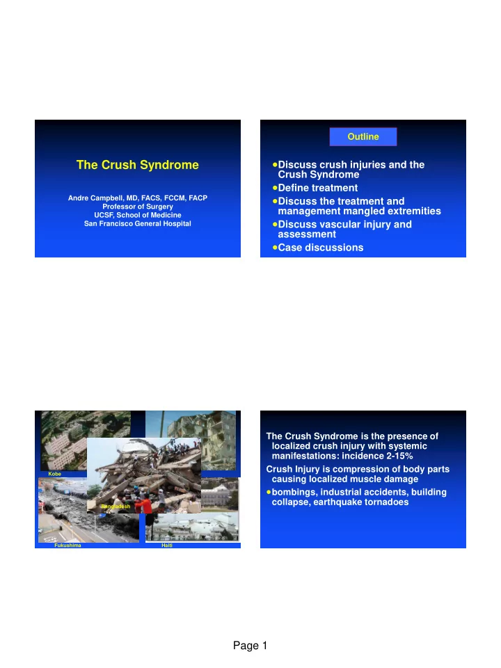
Kobe Armenia Fukushima Haiti Bangladesh
The Crush Syndrome is the presence of localized crush injury with systemic manifestations: incidence 2-15% Crush Injury is compression of body parts causing localized muscle damage
bombings, industrial accidents, building
collapse, earthquake tornadoes
SLIDE 2 Page 2
Crush Injury Muscle ischemia and Necrosis from Prolonged Pressure (Local effects)
Crush Syndrome
(Systemic Effects)
Fluid Retention in Extremities (third spacing) Hypotension Myoglobinuria Renal Failure Metabolic Abnormalities (electrolytes) Cardiac Arrhythmias Secondary Complications Compartment Syndrome
Crush Injuries
Injuries typically associated with disasters that
include muscle injury, renal failure and death
Man made-war and natural- earthquake Earthquakes 3-20% of crush injuries Building collapse up to 40% of extricated victims Vehicular Disaster Terrorist Acts- Oklahoma City, 9/11 Systemic manifestations of muscular cell damage
resulting from pressure of crushing
Crush Injuries
Recognized after the Messina earthquake of
1909 and during WWI by German MDs
First described in the English literature by
Bywaters and Beall in 1941 –Several patients who were crushed during WWII during the Blitz over London. –All patients died from renal failure despite resuscitation
Br Med J 1941;427-432
The Crush Syndrome
Characteristic Syndrome the results in rhadomyolysis, myoglobinuria, ARF.
Three criteria – Involvement of muscle mass – Prolonged compression 4-6 hrs. but can be < 1 hr – Compromised local circulation
Gonzalez, D Crit Care Med 2005 33. No 1(Suppl)
SLIDE 3
Page 3
Causes of Mortality after Untreated Crush Injury
Immediate:
–Severe head injury, traumatic asphyxia, and torso injuries
Early:
–Hyperkalemia, hypovolemic shock
Late:
–Renal failure, coagulopathy, and hemorrhage, sepsis
Clinical Manifestations Crush Syndrome
Hypotension:
–Massive 3rd spacing –Shock contributes to renal failure –Third spacing can lead to compartment syndrome
Renal Failure
–Rhadomyolysis releases myoglobin, K, P04, Cr, into circulation –Myoglobinuria leads to renal tubular necrosis –Release of electrolytes from ischemic muscle cause metabolic abnormality
Clinical Manifestations of the Crush Syndrome
Metabolic Abnormalities:
–Ca flow intracellularly through leaky membranes causing systemic hypocalcemia –K is released from muscle causing systemic hyperkalemia –Lactic Acid is released from ischemic muscle into systemic circulation, causing metabolic acidosis –Imbalance between K and Ca cause cardiac arrhythmias-acidosis makes it worst
SLIDE 4 Page 4
Clinical Manifestations
Electrolyte Disturbances
–Hyperkalemia, Hypocalcemia, Hyperphosphatemia, Metabolic acidosis
Renal
–Renal vasoconstriction due to shock –Pigment toxicity due to myoglobin –ATN –Luminal obstruction –Acute Renal Failure
Indicators of Severity
CPK elevation correlates with renal failure
(RF) and mortality
Risk of mortality and renal failure
increased with CK over 75,000 U/L
Other suggested counting limbs crushed
Crush one limb-RF 50%, two-RF-75%,
three RF- 100%
Oda J et al; J Trauma 1997;30:507-512
Crush Syndrome Pre-Hospital
Coordinate time of release with rescue
personnel
Mass casualty scenarios should be
discussed with personnel
Airway secured and protected from dust Adequate oxygenation Maintain body temperature Rapid transit to a trauma center Intravenous fluids, cardiac monitoring
Crush Syndrome Pre-hospital
Establish two large bore IVs Administer 1-2 liter or LR prior to extrication
–If prolonged infuse 1.5 liters/hr –Young and elderly be cautious about fluid
Sodium Bicarb 2 amps prior to extrication Cardiac monitoring Pain control PRN Extricate
SLIDE 5
Page 5
Definitive Treatment
Hypotensive:
–Massive fluid shifts –Hydration 1.5 liters/hour –Patient may gain massive amounts of weight in the resuscitation –Similar to Burn patients
Definitive Treatment
Renal Failure:
–Prevent renal failure with adequate hydration –Maintain diuresis of 300ml/hr with IV fluids and mannitol(carefully) –Triage to hemodialysis as needed »May need 60 days of Rx »Should return to normal function
Definitive Treatment
Metabolic Abnormalities:
–Acidosis: administer IV sodium bicarbonate until urine pH reaches 6.5 to prevent myoglobin deposition –Hyperkalemia/Hypocalcemia: administer Ca, sodium bicarbonate, insulin/D5W, consider kayexalate –Cardiac arrhythmias monitor for cardiac arrhythmias and arrest and treat
Definitive Treatment Secondary Complications
Monitor for compartment syndromes and do
fasciotomies as needed
Treat open wounds with antibiotics, tetanus
toxoid, and debridement
Monitor for pain, pallor, pulselessness,
paresthesias, paralysis- ischemia
Observe all crush injuries-even those who look
normal
Delays in hydration for more than 12 hours lead
to renal failure
Definitive surgery- amputations as needed
SLIDE 6 Page 6
J Trauma 2012;72:1626-1633 J Trauma 2012;72:1626-1633 J Trauma 2012;72:1626-1633
SLIDE 7 Page 7
Perte’s Syndrome or Traumatic Asphyxia
Craniocervical cyanosis Subconjunctival hemorrhage Multiple petechiae Neurological symptoms Results from sudden severe
compression of the thorax or upper abdomen or both
Valsalva is necessary before crush Associated injuries pulmonary
contusions, hemothorax or pneumothorax
Injury 2013;44:60-65
Aortic Crush Injuries vs. MVA Aortic injuries
Increase risk of Rhadomyolysis, ATN and
renal failure
Tendency to develop lower risk aortic
injuries than MVAs
Both type of patient must be followed
since they can progress
High rate of mortality in missed injuries
Injury 2013;44:60-65
SLIDE 8 Page 8
Retrospective Cohort design of data from
the NTDB 2007-2009
Assessed the result from 222 Level I & II
trauma centers of severely mangled extremities
1354 patients were analyzed and logistic
regression done to assess factors associated with amputations
21% of patients underwent amputations in
this study (9% early amputations)
J of Trauma 2013;74:597-603
Factors correlated with amputation
–Presence of severe head injury AIS>3 –Presence of shock in the ED(BP<90) –Limb injury type –High energy mechanism of injury –Age, comorbidities, and insurance status do not govern amputation rate –Injury type is the most important thing
J of Trauma 2013;74:597-603
Blunt Arterial Injury Salvage Rates
Have a high amputation rate
due to associated soft-tissue and nerve injuries (the mangled extremity)
These injuries may result in a
non-functional limb in spite of a successful revascularization
Blunt Vascular Trauma
Retrospective review at a Level I trauma center Jan 1995-Dec 2002 62 patients ISS>14.6, 93 vascular injuries,66% hard signs, 95% had
associated fracture
Age, ISS, and MESS was significantly different between
survivors and non-survivors
Injuries to the upper and lower extremity Shunt were used in 18 vessels prior to repair Anteroposterior tibia artery most commonly injured Amputation rate was 18% 3X that for penetrating injury
Rozycki G et al., J Trauma 2003;55:814-824
SLIDE 9 Page 9
Mangled Extremity
Indications for Primary
Amputation – Anatomically complete disruption of sciatic or posterior tibial nerves in adult even if vascular injury is repairable – Prolonged warm ischemia time – Life threatening sequelae » rhabdomyolysis
Mangled Extremity
Relative Indications for Primary
Amputation –Serious associated polytrauma –Severe ipsilateral foot trauma »loss of plantar skin/weight bearing surface –Anticipated protracted course to obtain soft-tissue coverage and skeletal reconstruction
Variables in Consideration of Limb Viability
Skin/Muscle Injury Bone Injury Ischemia (time, degree) Type of Vascular Injury Shock Age Infection Associated injuries (pulmonary, abdominal, head, etc.) Comorbid Disease (peripheral vascular disease, diabetes
mellitus, etc.)
Classification Systems
Mangled Extremity Syndrome Index (MESI)
– 9 variables
Predictive Salvage Index (PSI)
– 4 variables
Mangled Extremity Severity Score (MESS)
– 4 variables
Limb Salvage Index (LSI)
– 7 variables
NISSSA scoring system (Nerve Injury, Soft Tissue Injury, Skeletal
Injury, Shock, Age of Patient Score) – 6 variables
Hanover Fracture Scale(HFS)
– 12 variable
SLIDE 10 Page 10
Variable MESI PSI HFS LSI MESS NISSSA
Bone/FX type
+ + + + + +
Skin/muscle
+ + + + + +
Nerve
+
+
Vascular/ischemia
+ + + + + +
Contamination
+
+
+
Lag Time
+ + +
+
+
Comorbidity
+
- Smoking behavior
- Number of
Variables
9 4 12 7 4 6
Range
3-75 3-15 1-39 0-14 1-14 0-16
Cuttoff Point
20 8 15 6 7 9 Hoogendoorn, Werken, E J of Trauma 2002;28:1-10
J Trauma 2012;72:86-93
Vascular Injury – Non-invasive tests
“Soft signs”
large stable hematoma prior significant bleeding possible nerve damage proximity (<3cm, used to angio but only 10 – 20%
pos) Physical exam and API – arterial pressure index ABI <0.9, perform Duplex, color flow – to decide whether to angio, observe, OR Mandatory exploration not needed
Richardson, JD et al., Arch Surg 122:678, 1987
Vascular Injury – Non-invasive tests
“Soft signs” – large stable hematoma, prior
significant bleeding, possible nerve damage, proximity (<3cm, used to angio but
API – arterial pressure index (doppler sys
injured compared to non-injured) or ABI <0.9, Duplex, color flow – use to decide whether to angio
SLIDE 11 Page 11
Vascular Injury – Surg vs Angio
“Hard signs” – absent or dec distal pulse,
pulsatile bleeding, expanding hematoma, bruit
Depends on surgeon whether explore and
repair or request angio – usually do study first if multiple (gsw), blunt trauma (pre-
- rtho), or possible vasc intervention
Vascular Injury – Surg vs Angio
“Hard signs”
absent or decreased distal pulse-no brainer
usually
pulsatile bleeding expanding hematoma bruit
Depends on surgeon’s experience whether explore and repair or request angio, study requested for penetrating (gsw), or possible vascular intervention
Patient with suspected vascular injury
Resuscitation “Hard” signs Soft signs API>0.9 API<0.9 Observe Duplex AGRAM Clinical Follow-up Positive Negative Surgical Exploration AGRAM Clinical Follow-up Positive
Mattox, Feliciano, Moore, Fourth Edition , 2000
Temporary Vascular Shunt
SLIDE 12
Page 12
Definitive Vascular Repair Mechanisms of Arterial Injury
Patient experiences – blunt, penetrating or
combined mechanism (fx/dislocation)
Artery experiences – crush, shear, stretch,
laceration, shock wave
Artery responses – spasm, tear (partial or
complete transection), bleeding with hematoma, intimal injury, thrombosis
Angio shows – narrowing (spasm or extrinsic
compression), intimal injury (flap, dissection), extravasation, pseudoaneurysm, AVF, abrupt cut-off, intraluminal thrombus Train Accident
SLIDE 13
Page 13
Iraq IED Iraq IED
Mangled Extremity Management
Involves a determination of both the
feasibility (restoring viability) and advisability (restoring function) of salvaging the limb
Should be a coordinated effort of the
trauma, orthopedic, vascular and plastic surgeons starting at the initial evaluation of the patient
SLIDE 14
Page 14
Release compartments Reduce dislocations Debride open wounds Stabilize long bones Restore vascular flow
Goal of Early Management of Extremity Injury in the Polytrauma Patient: Stop the Ongoing Injury
Crush Injury Signs and Symptoms- Summary
Compression in excess of 60 minutes Involvement of large muscle mass Absent pulse and capillary refill return to
distal limb
Pale, clammy, cool skin Usually absence of pain in affect region Onset of shock Consider prior to extraction
The Crush Syndrome Treatment Summary
Radical Surgery Fluid resuscitation Alkalinization Mannitol Diuresis Compartment Syndromes must be
treated –Pain, Pallor, Pulselessnes, Parathesias and Paralysis
Conclusions
Difficult situations in patients with multiple
injuries
Patients with crush injuries are complex Have to make a decision early if it is a
salvageable extremity
Amputation takes courage Multidisciplinary approach is the best when
the patient initially presents